

Fluorescence Lifetime Changes Induced by Laser Irradiation: A Preclinical Study towards the Evaluation of Retinal Metabolic States
- PMID: 34199212
- PMCID: PMC8231852
- DOI: 10.3390/life11060555
Fluorescence Lifetime Changes Induced by Laser Irradiation: A Preclinical Study towards the Evaluation of Retinal Metabolic States
Abstract
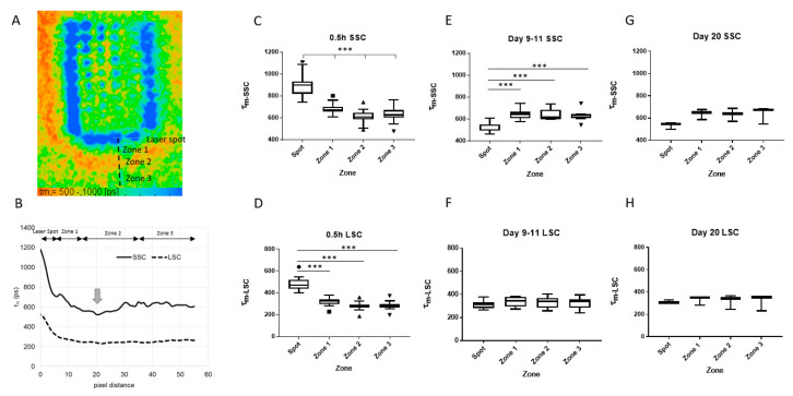
Fluorescence Lifetime (FLT) of intrinsic fluorophores may alter under the change in metabolic state. In this study, the FLT of rabbit retina was investigated in vivo after laser irradiation using fluorescence lifetime imaging ophthalmoscopy (FLIO). The retina of the Chinchilla bastard rabbits was irradiated with a 514 nm diode laser. FLIO, fundus photography, and optical coherence tomography (OCT) were conducted 30 min and 1 to 3 weeks after treatment. After strong coagulation, the FLT at laser spots was significantly elongated immediately after irradiation, conversely shortened after more than a week. Histological examination showed eosinophilic substance and melanin clumping in subretinal space at the coagulation spots older than one week. The FLT was also elongated right around the coagulation spots, which corresponded to the discontinuous ellipsoid zone (EZ) on OCT. This EZ change was recovered after one week, and the FLT became the same level as the surroundings. In addition, there was a region around the laser spot where the FLT was temporarily shorter than the surrounding area. When weak pulse energy was applied to selectively destroy only the RPE, a shortening of the FLT was observed immediately around the laser spot within one week after irradiation. FLIO could serve as a tool to evaluate the structural and metabolic response of the retina to laser treatments.
Keywords: fluorescence lifetime imaging ophthalmoscopy; metabolic change; retinal laser treatment.
Conflict of interest statement
The authors declare no conflict of interest.
Figures


Similar articles
-
Detection sensitivity of fluorescence lifetime imaging ophthalmoscopy for laser-induced selective damage of retinal pigment epithelium.Graefes Arch Clin Exp Ophthalmol. 2024 Sep;262(9):2885-2895. doi: 10.1007/s00417-024-06449-2. Epub 2024 Apr 8. Graefes Arch Clin Exp Ophthalmol. 2024. PMID: 38587656 Free PMC article.
-
Impact of cigarette smoking on fluorescence lifetime of ocular fundus.Sci Rep. 2023 Jul 17;13(1):11484. doi: 10.1038/s41598-023-37484-4. Sci Rep. 2023. PMID: 37460627 Free PMC article.
-
Fluorescence Lifetime Imaging Ophthalmoscopy of the Retinal Pigment Epithelium During Wound Healing After Laser Irradiation.Transl Vis Sci Technol. 2019 Sep 18;8(5):12. doi: 10.1167/tvst.8.5.12. eCollection 2019 Sep. Transl Vis Sci Technol. 2019. PMID: 31588376 Free PMC article.
-
Fluorescence Lifetime Imaging Ophthalmoscopy (FLIO).2019 Aug 14. In: Bille JF, editor. High Resolution Imaging in Microscopy and Ophthalmology: New Frontiers in Biomedical Optics [Internet]. Cham (CH): Springer; 2019. Chapter 10. 2019 Aug 14. In: Bille JF, editor. High Resolution Imaging in Microscopy and Ophthalmology: New Frontiers in Biomedical Optics [Internet]. Cham (CH): Springer; 2019. Chapter 10. PMID: 32091850 Free Books & Documents. Review.
-
[Pathophysiology of macular diseases--morphology and function].Nippon Ganka Gakkai Zasshi. 2011 Mar;115(3):238-74; discussion 275. Nippon Ganka Gakkai Zasshi. 2011. PMID: 21476310 Review. Japanese.
Cited by
-
Retinal Disease and Metabolism.Life (Basel). 2022 Jan 27;12(2):183. doi: 10.3390/life12020183. Life (Basel). 2022. PMID: 35207471 Free PMC article.
-
Artificial Intelligence in Fluorescence Lifetime Imaging Ophthalmoscopy (FLIO) Data Analysis-Toward Retinal Metabolic Diagnostics.Diagnostics (Basel). 2024 Feb 16;14(4):431. doi: 10.3390/diagnostics14040431. Diagnostics (Basel). 2024. PMID: 38396470 Free PMC article.
-
Detection sensitivity of fluorescence lifetime imaging ophthalmoscopy for laser-induced selective damage of retinal pigment epithelium.Graefes Arch Clin Exp Ophthalmol. 2024 Sep;262(9):2885-2895. doi: 10.1007/s00417-024-06449-2. Epub 2024 Apr 8. Graefes Arch Clin Exp Ophthalmol. 2024. PMID: 38587656 Free PMC article.
-
Impact of cigarette smoking on fluorescence lifetime of ocular fundus.Sci Rep. 2023 Jul 17;13(1):11484. doi: 10.1038/s41598-023-37484-4. Sci Rep. 2023. PMID: 37460627 Free PMC article.
References
-
- Brinkmann R., Roider J., Birngruber R. Selective retina therapy (SRT): A review on methods, techniques, preclinical and first clinical results. Bull. Soc. Belge Ophtalmol. 2006;302:51–69. - PubMed
Grants and funding
LinkOut - more resources
Full Text Sources
