. 2021 Jun 13;22(12):6339.
doi: 10.3390/ijms22126339.

n-Butylidenephthalide Modulates Autophagy to Ameliorate Neuropathological Progress of Spinocerebellar Ataxia Type 3 through mTOR Pathway

Affiliations

n-Butylidenephthalide Modulates Autophagy to Ameliorate Neuropathological Progress of Spinocerebellar Ataxia Type 3 through mTOR Pathway

Jui-Hao Lee et al. Int J Mol Sci. .

Abstract

Spinocerebellar ataxia type 3 (SCA3), a hereditary and lethal neurodegenerative disease, is attributed to the abnormal accumulation of undegradable polyglutamine (polyQ), which is encoded by mutated ataxin-3 gene (ATXN3). The toxic fragments processed from mutant ATXN3 can induce neuronal death, leading to the muscular incoordination of the human body. Some treatment strategies of SCA3 are preferentially focused on depleting the abnormal aggregates, which led to the discovery of small molecule n-butylidenephthalide (n-BP). n-BP-promoted autophagy protected the loss of Purkinje cell in the cerebellum that regulates the network associated with motor functions. We report that the n-BP treatment may be effective in treating SCA3 disease. n-BP treatment led to the depletion of mutant ATXN3 with the expanded polyQ chain and the toxic fragments resulting in increased metabolic activity and alleviated atrophy of SCA3 murine cerebellum. Furthermore, n-BP treated animal and HEK-293GFP-ATXN3-84Q cell models could consistently show the depletion of aggregates through mTOR inhibition. With its unique mechanism, the two autophagic inhibitors Bafilomycin A1 and wortmannin could halt the n-BP-induced elimination of aggregates. Collectively, n-BP shows promising results for the treatment of SCA3.

Keywords: MJD; PolyQ; Purkinje cell; SCA3; atxain-3; autophagy; mTOR; toxic fragment.


Conflict of interest statement

The authors declare no conflict of interest.

Figures

Figure 1
Figure 1
Administration of n-BP significantly alleviated impairments of motor coordination and ameliorated gait/locomotion in the SCA3 mouse model. (A) The representative designed study showing 23-week-old wild-type and SCA3 mice were divided into four groups (each n = 10 at the beginning): WT, SCA3, vehicle, or n-BP (50 mg/kg b.i.d.) treated with SCA3 (SCA3-V or SCA3-BP) for 5 weeks. Behavior studies were performed every week, and the studied animals were euthanized at first, third, and fifth weeks for sample collection and indicated analysis. (B) A significantly improved motor coordination of the n-BP treated SCA3 mice in accelerating (0–40 r.p.m. within 5 min) rotarod test was recorded. The n-BP treated SCA3 mice had better motor performance and balance compared to SCA3’s non-treated and vehicle groups. Asterisk presents the calculated p value of SCA3-BP versus WT or SCA3-V. To figure out the best response to n-BP during the treatment, small groups of 1–2 mice were additionally prepared for the behavioral evaluation at the sixth week. (C,D) Representative images of footprint patterns displaying the gait of n-BP administered SCA3 mice were similar to WT animals at the fifth week (28-week-old mice). Based on the 5 weeks’ footprinting test, SCA3-BP group presented improved coordination with shorter footprint overlap and longer stride length than vehicle or non-treated animals. Moreover, the long front and the hind base width in SCA3 and SCA3-V groups were presented. The 5 weeks of n-BP treatment made a significant improvement on balance of SCA3 mice (†: **, ‡: ***, #: ****). Animal number for the statistics was indicated as n. Data represent mean ± s.d.; * p < 0.05, ** p < 0.01, *** p < 0.001, **** p < 0.0001 (Student’s t-test).
Figure 1
Figure 1
Administration of n-BP significantly alleviated impairments of motor coordination and ameliorated gait/locomotion in the SCA3 mouse model. (A) The representative designed study showing 23-week-old wild-type and SCA3 mice were divided into four groups (each n = 10 at the beginning): WT, SCA3, vehicle, or n-BP (50 mg/kg b.i.d.) treated with SCA3 (SCA3-V or SCA3-BP) for 5 weeks. Behavior studies were performed every week, and the studied animals were euthanized at first, third, and fifth weeks for sample collection and indicated analysis. (B) A significantly improved motor coordination of the n-BP treated SCA3 mice in accelerating (0–40 r.p.m. within 5 min) rotarod test was recorded. The n-BP treated SCA3 mice had better motor performance and balance compared to SCA3’s non-treated and vehicle groups. Asterisk presents the calculated p value of SCA3-BP versus WT or SCA3-V. To figure out the best response to n-BP during the treatment, small groups of 1–2 mice were additionally prepared for the behavioral evaluation at the sixth week. (C,D) Representative images of footprint patterns displaying the gait of n-BP administered SCA3 mice were similar to WT animals at the fifth week (28-week-old mice). Based on the 5 weeks’ footprinting test, SCA3-BP group presented improved coordination with shorter footprint overlap and longer stride length than vehicle or non-treated animals. Moreover, the long front and the hind base width in SCA3 and SCA3-V groups were presented. The 5 weeks of n-BP treatment made a significant improvement on balance of SCA3 mice (†: **, ‡: ***, #: ****). Animal number for the statistics was indicated as n. Data represent mean ± s.d.; * p < 0.05, ** p < 0.01, *** p < 0.001, **** p < 0.0001 (Student’s t-test).
Figure 2
Figure 2
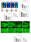
n-BP protected the Purkinje cells from loss to increase the metabolic activity in the cerebellum in the SCA3 mice. (A) The representative PET ([18F]FDG) and MR images, in coronal (C), sagittal (S), and transverse (T) views, of SCA3 mice showing that n-BP treatment for 12 weeks could alleviate the impairment of glucose analogue metabolism and restore the cerebellar function (yellow ROI). Each group contained three 35-week-old mice for the analysis. (B) The panel presents the values corresponding to the standard uptake value (SUV) based on the in vivo imaging of glucose metabolic activity (yellow ROI of Figure 2A). (C) Histogram indicated the MR evaluated cerebellar size in voxel, reflecting the cerebellar atrophy in SCA3 or SCA3-V animals. (D) The images exhibit the whole brain of WT and SCA3 mice with indicated treatments for five weeks. Statistical results suggested that the n-BP treatment could mitigate the atrophy of cerebellum, and no difference in cerebellum size (x × y = cm × cm) was found between WT and SCA3-BP groups (at 28 weeks of age). Three animals were used for the statistical data. (E) Cerebellar tissue sections of the mice were stained with calbindin (green, Alexa Fluor 488 probed) to label the Purkinje cells in cerebellar lobes III–V. The representative images and zoomed parts of Purkinje cells are shown. Purkinje cells without fragmented neurites are indicated by arrows. The counted number of all Purkinje cells in each captured field are calculated in the bottom panel. Bar = 50 μm. Data represent mean ± s.d. of three independent experiments (total cell counts in 28 fields). * p < 0.05, ** p < 0.01, *** p < 0.001, **** p < 0.0001 (Student’s t-test).
Figure 3
Figure 3
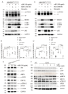
n-BP reduced mutant ATXN3 aggregates and toxic fragments through promoting autophagy in vitro and in vivo. The ATXN3 aggregates (A) and the polyQ proteins (B) (red, Alexa Fluor 594 conjugated) were located at the Purkinje cells (green, Alexa Fluor 488 labeled) in the cerebellum tissues from 28-week-old mice. Hoechst 33342 was used to label nuclei. Arrows mark the colocalization of ATXN3/polyQ and calbindin in the zoomed images. Bar = 50 μm. (C) Each sampling cerebellum was analyzed to identify the expression of mutated ATXN3 (m-ATXN3) with polyQ expansion (polyQ) and toxic fragments (TFs) by Western blotting assay. n-BP administration could efficiently decrease the accumulation of these abnormal proteins versus vehicle or non-treated SCA3 mice. Arrow indicates the wild-type ATXN3 at 45 kD. (D,E) The representative Western blot analysis showing the increased level of BECN1, LC3B-II, ATG3, and ATG7 and a concurrent decreased expression of p62 detected in the murine cerebellum from WT and SCA3-BP groups. Anti-LC3B, p62, and GAPDH antibodies were used on the same blot. The ratios of LC3B-II and p62 to GAPDH were calculated using ImageJ software and normalized to WT. (F,G) HEK-293 cells transfected with GFP-ATXN3-84Q or control GFP-ATXN3-28Q were treated with n-BP (50 or 100 μg/mL) or vehicle (0 μg/mL). After 6 h treatment, cell samples were subjected to the immunoblotting assay. Findings suggested that n-BP could reduce the accumulated m-ATXN3 and TFs and the expression of p62 in HEK-293GFP-ATXN3-84Q cell models. Consistently, expression of autophagic players including BECN1, LC3B-II, ATG3, and ATG7 could be induced by n-BP. GAPDH, LC3B-II, and p62 were detected on the same blot. The ratios of LC3B-II and p62 to GAPDH were calculated and respectively normalized to control or HEK-293GFP-ATXN3-84Q cells. GAPDH was used as an internal control. Data represent mean ± s.d.; * p < 0.05, ** p < 0.01, *** p < 0.001 (Student’s t-test). All experiments were repeated at least three times.
Figure 3
Figure 3
n-BP reduced mutant ATXN3 aggregates and toxic fragments through promoting autophagy in vitro and in vivo. The ATXN3 aggregates (A) and the polyQ proteins (B) (red, Alexa Fluor 594 conjugated) were located at the Purkinje cells (green, Alexa Fluor 488 labeled) in the cerebellum tissues from 28-week-old mice. Hoechst 33342 was used to label nuclei. Arrows mark the colocalization of ATXN3/polyQ and calbindin in the zoomed images. Bar = 50 μm. (C) Each sampling cerebellum was analyzed to identify the expression of mutated ATXN3 (m-ATXN3) with polyQ expansion (polyQ) and toxic fragments (TFs) by Western blotting assay. n-BP administration could efficiently decrease the accumulation of these abnormal proteins versus vehicle or non-treated SCA3 mice. Arrow indicates the wild-type ATXN3 at 45 kD. (D,E) The representative Western blot analysis showing the increased level of BECN1, LC3B-II, ATG3, and ATG7 and a concurrent decreased expression of p62 detected in the murine cerebellum from WT and SCA3-BP groups. Anti-LC3B, p62, and GAPDH antibodies were used on the same blot. The ratios of LC3B-II and p62 to GAPDH were calculated using ImageJ software and normalized to WT. (F,G) HEK-293 cells transfected with GFP-ATXN3-84Q or control GFP-ATXN3-28Q were treated with n-BP (50 or 100 μg/mL) or vehicle (0 μg/mL). After 6 h treatment, cell samples were subjected to the immunoblotting assay. Findings suggested that n-BP could reduce the accumulated m-ATXN3 and TFs and the expression of p62 in HEK-293GFP-ATXN3-84Q cell models. Consistently, expression of autophagic players including BECN1, LC3B-II, ATG3, and ATG7 could be induced by n-BP. GAPDH, LC3B-II, and p62 were detected on the same blot. The ratios of LC3B-II and p62 to GAPDH were calculated and respectively normalized to control or HEK-293GFP-ATXN3-84Q cells. GAPDH was used as an internal control. Data represent mean ± s.d.; * p < 0.05, ** p < 0.01, *** p < 0.001 (Student’s t-test). All experiments were repeated at least three times.
Figure 4
Figure 4
n-BP acted in early stage of autophagy to potentiate the elimination of aggregates through inhibiting mTOR pathway. HEK-293GFP-ATXN3-84Q cell model was used to dissect the mechanistic action of n-BP under Bafilomycin A1 (BafA1) (A) or wortmannin (Wort) (C) exposures. n-BP or starvation inducing BECN1, LC3B-II, and ATG7 expression and p62 degradation and reducing m-ATXN3 and TF levels indicated the activation of autophagy. (A,B) Combined treatment of n-BP and BafA1 could not result in the reduction of m-ATXN3 and TFs. Autophagic flux, revealed by ratios of LC3B-II and p62 to GAPDH, was measured, and the changes provided the effect of n-BP on autophagic promotion. (C,D) In addition, wortmannin (Wort) could inhibit the n-BP-induced autophagic process and retain mutant ATXN3 proteins in the cells. The levels of LC3B-II, p62, and ATG7 were not different from vehicle control and Wort with or without n-BP treatment. (B,D) Ratios of LC3B-II and p62 to GAPDH were calculated and normalized to the vehicle control. GAPDH, LC3B, and p62 were detected on the same blot. (E) In HEK-293GFP-ATXN3-84Q cells, Western blot results showed that n-BP (50 or 100 μg/mL) could reduce the level of phosphorylated AKT (p-AKT) and ERK1/2 (p-ERK1/2) and induce the level of phosphorylated AMPK (p-AMPK), respectively. Consequently, p-mTOR was detected in a lower level than vehicle control (0 μg/mL). (F) Cerebellar tissues were sampled from WT, SCA3-BP, and vehicle or non-treated SCA3 groups (28-week-old mice) for Western blotting analysis. The expression levels of phosphorylated AKT, ERK1/2, and mTOR were lower in WT and n-BP treated SCA3 mice compared to SCA3 and SCA3-V groups. AMPK activation was also detected in SCA3-BP group. GAPDH was used as an internal control. Data represent mean ± s.d.; * p < 0.05, ** p < 0.01, *** p < 0.001 (Student’s t-test). All experiments were repeated at least three times.

Similar articles

Cited by

References

    1. Strong T.V., Tagle D.A., Valdes J.M., Elmer L.W., Boehm K., Swaroop M., Kaatz K.W., Collins F.S., Albin R.L. Widespread expression of the human and rat Huntington′s disease gene in brain and nonneural tissues. Nat. Genet. 1993;5:259–265. doi: 10.1038/ng1193-259. - DOI - PubMed
    1. Kawaguchi Y., Okamoto T., Taniwaki M., Aizawa M., Inoue M., Katayama S., Kawakami H., Nakamura S., Nishimura M., Akiguchi I. CAG expansions in a novel gene for Machado-Joseph disease at chromosome 14q32. 1. Nat. Genet. 1994;8:221–228. doi: 10.1038/ng1194-221. - DOI - PubMed
    1. Koide R., Ikeuchi T., Onodera O., Tanaka H., Igarashi S., Endo K., Takahashi H., Kondo R., Ishikawa A., Hayashi T. Unstable expansion of CAG repeat in hereditary dentatorubral—Pallidoluysian atrophy (DRPLA) Nat. Genet. 1994;6:9–13. doi: 10.1038/ng0194-9. - DOI - PubMed
    1. David G., Abbas N., Stevanin G., Dürr A., Yvert G., Cancel G., Weber C., Imbert G., Saudou F., Antoniou E. Cloning of the SCA7 gene reveals a highly unstable CAG repeat expansion. Nat. Genet. 1997;17:65–70. doi: 10.1038/ng0997-65. - DOI - PubMed
    1. Zhuchenko O., Bailey J., Bonnen P., Ashizawa T., Stockton D.W., Amos C., Dobyns W.B., Subramony S., Zoghbi H.Y., Lee C.C. Autosomal dominant cerebellar ataxia (SCA6) associated with small polyglutamine expansions in the α 1A-voltage-dependent calcium channel. Nat. Genet. 1997;15:62–69. doi: 10.1038/ng0197-62. - DOI - PubMed

MeSH terms

LinkOut - more resources