. 2021 Jun 2;13(11):2760.
doi: 10.3390/cancers13112760.

Follow-Up Study Confirms the Presence of Gastric Cancer DNA Methylation Hallmarks in High-Risk Precursor Lesions

Affiliations

Follow-Up Study Confirms the Presence of Gastric Cancer DNA Methylation Hallmarks in High-Risk Precursor Lesions

Antonio Gómez et al. Cancers (Basel). .

Abstract

To adopt prevention strategies in gastric cancer, it is imperative to develop robust biomarkers with acceptable costs and feasibility in clinical practice to stratified populations according to risk scores. With this aim, we applied an unbiased genome-wide CpG methylation approach to a discovery cohort composed of gastric cancer (n = 24), and non-malignant precursor lesions (n = 64). Then, candidate-methylation approaches were performed in a validation cohort of precursor lesions obtained from an observational longitudinal study (n = 264), with a 12-year follow-up to identify repression or progression cases. H. pylori stratification and histology were considered to determine their influence on the methylation dynamics. As a result, we ascertained that intestinal metaplasia partially recapitulates patterns of aberrant methylation of intestinal type of gastric cancer, independently of the H. pylori status. Two epigenetically regulated genes in cancer, RPRM and ZNF793, consistently showed increased methylation in intestinal metaplasia with respect to earlier precursor lesions. In summary, our result supports the need to investigate the practical utilities of the quantification of DNA methylation in candidate genes as a marker for disease progression. In addition, the H. pylori-dependent methylation in intestinal metaplasia suggests that pharmacological treatments aimed at H. pylori eradication in the late stages of precursor lesions do not prevent epigenome reprogramming toward a cancer signature.

Keywords: CpG methylation; Helicobacter pylori; cancer risk prediction; intestinal type of gastric cancer; precursor lesions.


Conflict of interest statement

The authors declare no conflict of interest.

Figures

Figure 1
Figure 1
CpG methylation signature of intestinal and diffuse types of gastric cancer. (A) Center, workflow of the cancer-related study. Circus graphs for genome-wide DNA methylation levels in diffuse (right) and intestinal (left) gastric tumors. The inner track indicates the β-value of the non-tumoral and the outer circle represents the cancer sample. (B) Total number of hypermethylation and hypomethylation events in gastric cancer, relative to adjacent non-tumoral samples. (C) Genomic distribution of differentially methylated CpGs in the intestinal and diffuse types of gastric cancer.
Figure 2
Figure 2
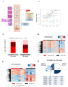
CpG methylation signature of the gastric precancerous cascade. (A) Schematic representation of the precursor lesion pathway. Two genome-wide CpG methylation analyses were performed: (1) H. pylori-related study to identify the timing of epigenetic aberrations due to bacterial infection, and (2) a study of the hypermethylation events observed in IM that could act as biomarkers of cancer progression. (B) Boxplots showing overall β-values for NM, NAG, CAG, IM and GC samples. Overall beta values were calculated as the mean of all CpGs in each sample, then plotted as boxplots for all groups. Each box represents the IQR with horizontal lines representing the median. Whiskers are extended to within 1.5 IQR of the upper and lower quantities. Data points falling outside this range are displayed independently. The p-values were calculated using Student’s t-test. (C) Total number of hypermethylation and hypomethylation events in IM relative to normal tissues. (D) Supervised clustering of methylation values in the IM samples stratified by H. pylori infection. (E) Supervised clustering of methylation values in the CAG samples considering the presence or absence of H. pylori. (F) Overlapped hypermethylation in IM and intestinal type of gastric cancer. CAG, multifocal chronic atrophic gastritis; GC, gastric cancer; IM, intestinal metaplasia; NAG, non-atrophic gastritis; NM, normal mucosa.
Figure 3
Figure 3
Methylation of ZNF793 and RPRM genes in intestinal type of gastric cancer and precursor lesions. (A) Methylation level of ZNF793 and RPRM genes in intestinal type of gastric cancer. Values from 13 adjacent-non-tumoral samples paired with the tumor sample are shown. Methylation values represent the average of β-value obtained for the three CG probes in the methylation array (paired t-test, two-tailed, p < 0.05, * p-value ≤ 0.05, *** p-value ≤ 0.001). (B) CpG methylation at RPRM and ZNF793 promoter in three anatomic intestinal regions (antrum, cardia and body) of intestinal metaplasia patients and normal mucosae (available data from GSE103186).
Figure 4
Figure 4
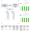
Methylation of ZNF793 and RPRM genes in a 12-year follow-up study of gastric cancer precursor lesions. (A) Characteristics of the validation cohort of 264 samples corresponding to precursor lesions obtained from an observational longitudinal study. (B) Methylation events for ZNF793 (up) and RPRM (down) genes in precursor lesions. (C) Association analysis between the progression in the cascade of gastric precursor lesions and the methylation of ZNF793 and RPRM genes. The p-values are obtained from Fisher’s exact test. (D) Number of cases of progression to gastric cancer, after a 12-year follow-up, for gastric precursor lesions. 1 Methylation state of ZNF793 and RPRM promoters at the recruitment biopsy are indicated. CAG, multifocal chronic atrophic gastritis; CG, gastric cancer; D, dysplasia; IM, intestinal metaplasia; NAG, non-atrophic gastritis; NM, normal mucosa; * p-value ≤ 0.05, ** p-value ≤ 0.01.

References

    1. Ferlay J., Soerjomataram I., Dikshit R., Eser S., Mathers C., Rebelo M., Parkin D.M., Forman D., Bray F. Cancer incidence and mortality worldwide: Sources, methods and major patterns in GLOBOCAN 2012. Int. J. Cancer. 2015;136:E359–E386. doi: 10.1002/ijc.29210. - DOI - PubMed
    1. Shi Y., Zhou Y. The role of surgery in the treatment of gastric cancer. J. Surg. Oncol. 2010;101:687–692. doi: 10.1002/jso.21455. - DOI - PubMed
    1. González C.A., Sanz-Anquela J.M., Gisbert J.P., Correa P. Utility of subtyping intestinal metaplasia as marker of gastric cancer risk. A review of the evidence. Int. J. Cancer. 2013;133:1023–1032. doi: 10.1002/ijc.28003. - DOI - PMC - PubMed
    1. Lauren P. The two histological main types of gastric carcinoma: Diffuse and so-called intestinal-type carcinoma. An attempt at a histo-clinical classification. Acta Pathol. Microbiol. Scand. 1965;64:31–49. doi: 10.1111/apm.1965.64.1.31. - DOI - PubMed
    1. Correa P., Piazuelo M.B. The gastric precancerous cascade. J. Dig. Dis. 2012;13:2–9. doi: 10.1111/j.1751-2980.2011.00550.x. - DOI - PMC - PubMed