

A Tissue-Engineered Tracheobronchial In Vitro Co-Culture Model for Determining Epithelial Toxicological and Inflammatory Responses
- PMID: 34199462
- PMCID: PMC8226664
- DOI: 10.3390/biomedicines9060631
A Tissue-Engineered Tracheobronchial In Vitro Co-Culture Model for Determining Epithelial Toxicological and Inflammatory Responses
Abstract
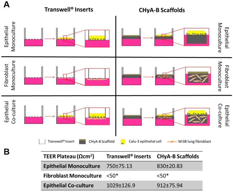
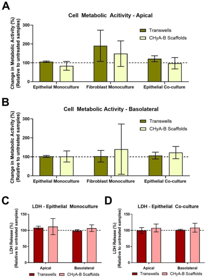
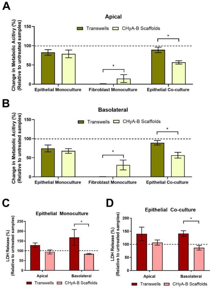
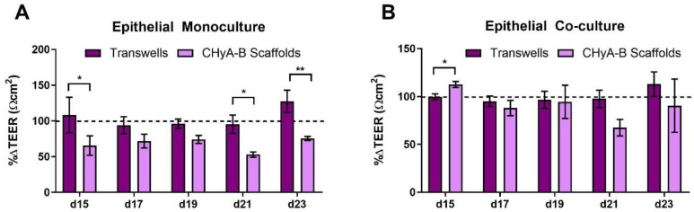
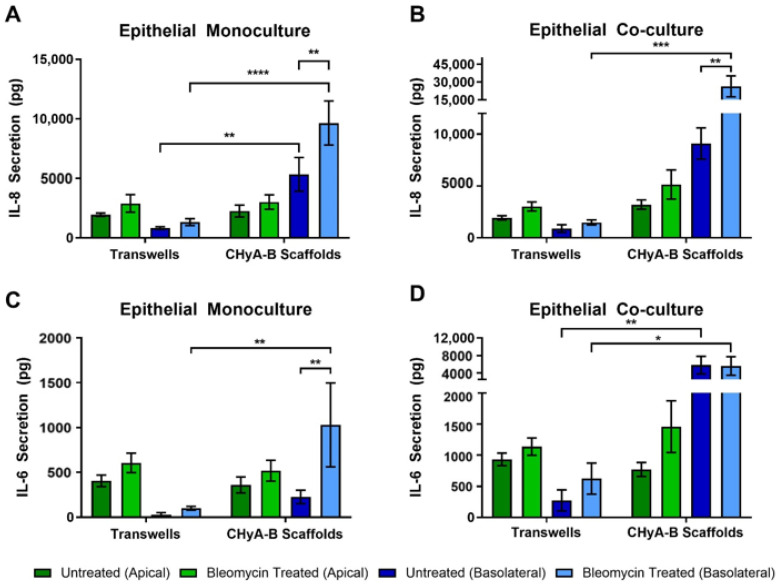
Translation of novel inhalable therapies for respiratory diseases is hampered due to the lack of in vitro cell models that reflect the complexity of native tissue, resulting in many novel drugs and formulations failing to progress beyond preclinical assessments. The development of physiologically-representative tracheobronchial tissue analogues has the potential to improve the translation of new treatments by more accurately reflecting in vivo respiratory pharmacological and toxicological responses. Herein, advanced tissue-engineered collagen hyaluronic acid bilayered scaffolds (CHyA-B) previously developed within our group were used to evaluate bacterial and drug-induced toxicity and inflammation for the first time. Calu-3 bronchial epithelial cells and Wi38 lung fibroblasts were grown on either CHyA-B scaffolds (3D) or Transwell® inserts (2D) under air liquid interface (ALI) conditions. Toxicological and inflammatory responses from epithelial monocultures and co-cultures grown in 2D or 3D were compared, using lipopolysaccharide (LPS) and bleomycin challenges to induce bacterial and drug responses in vitro. The 3D in vitro model exhibited significant epithelial barrier formation that was maintained upon introduction of co-culture conditions. Barrier integrity showed differential recovery in CHyA-B and Transwell® epithelial cultures. Basolateral secretion of pro-inflammatory cytokines to bacterial challenge was found to be higher from cells grown in 3D compared to 2D. In addition, higher cytotoxicity and increased basolateral levels of cytokines were detected when epithelial cultures grown in 3D were challenged with bleomycin. CHyA-B scaffolds support the growth and differentiation of bronchial epithelial cells in a 3D co-culture model with different transepithelial resistance in comparison to the same co-cultures grown on Transwell® inserts. Epithelial cultures in an extracellular matrix like environment show distinct responses in cytokine release and metabolic activity compared to 2D polarised models, which better mimic in vivo response to toxic and inflammatory stimuli offering an innovative in vitro platform for respiratory drug development.
Keywords: 3D in vitro models; air-liquid interface; bleomycin; co-culture; collagen; epithelium; lipopolysaccharide; respiratory tissue engineering; toxicology.
Conflict of interest statement
The authors declare no conflict of interest. The funders had no role in the design of the study; in the collection, analyses, or interpretation of data; in the writing of the manuscript, or in the decision to publish the results.
Figures

Similar articles
-
The development of a tissue-engineered tracheobronchial epithelial model using a bilayered collagen-hyaluronate scaffold.Biomaterials. 2016 Apr;85:111-27. doi: 10.1016/j.biomaterials.2016.01.065. Epub 2016 Feb 1. Biomaterials. 2016. PMID: 26871888
-
Triple co-culture of human alveolar epithelium, endothelium and macrophages for studying the interaction of nanocarriers with the air-blood barrier.Acta Biomater. 2019 Jun;91:235-247. doi: 10.1016/j.actbio.2019.04.037. Epub 2019 Apr 18. Acta Biomater. 2019. PMID: 31004840
-
Development of biomimetic co-culture and tri-culture models to mimic the complex structure of the alveolar-capillary barrier.Biomater Adv. 2023 Nov;154:213620. doi: 10.1016/j.bioadv.2023.213620. Epub 2023 Sep 8. Biomater Adv. 2023. PMID: 37690344
-
3D tissue-engineered lung models to study immune responses following viral infections of the small airways.Stem Cell Res Ther. 2022 Sep 7;13(1):464. doi: 10.1186/s13287-022-03134-1. Stem Cell Res Ther. 2022. PMID: 36071442 Free PMC article. Review.
-
Air-liquid interface (ALI) impact on different respiratory cell cultures.Eur J Pharm Biopharm. 2023 Mar;184:62-82. doi: 10.1016/j.ejpb.2023.01.013. Epub 2023 Jan 22. Eur J Pharm Biopharm. 2023. PMID: 36696943 Review.
Cited by
-
A miniaturized multicellular platform to mimic the 3D structure of the alveolar-capillary barrier.Front Bioeng Biotechnol. 2024 Apr 5;12:1346660. doi: 10.3389/fbioe.2024.1346660. eCollection 2024. Front Bioeng Biotechnol. 2024. PMID: 38646009 Free PMC article.
-
Advancements in lung regeneration: from bench to bedside.J Transl Med. 2025 Feb 4;23(1):154. doi: 10.1186/s12967-024-05954-6. J Transl Med. 2025. PMID: 39905476 Free PMC article. Review.
-
Interaction of Neisseria meningitidis carrier and disease isolates of MenB cc32 and MenW cc22 with epithelial cells of the nasopharyngeal barrier.Front Cell Infect Microbiol. 2024 May 2;14:1389527. doi: 10.3389/fcimb.2024.1389527. eCollection 2024. Front Cell Infect Microbiol. 2024. PMID: 38756230 Free PMC article.
References
-
- Chang L.H., Rivera M.P. Respiratory diseases: Meeting the challenges of screening, prevention, and treatment. N. Carol. Med. J. 2013;74:385–392. - PubMed
Grants and funding
LinkOut - more resources
Full Text Sources
