Review
. 2021 Jun 4;22(11):6062.
doi: 10.3390/ijms22116062.

Minor Intron Splicing from Basic Science to Disease

Affiliations
Review

Minor Intron Splicing from Basic Science to Disease

Ettaib El Marabti et al. Int J Mol Sci. .

Abstract

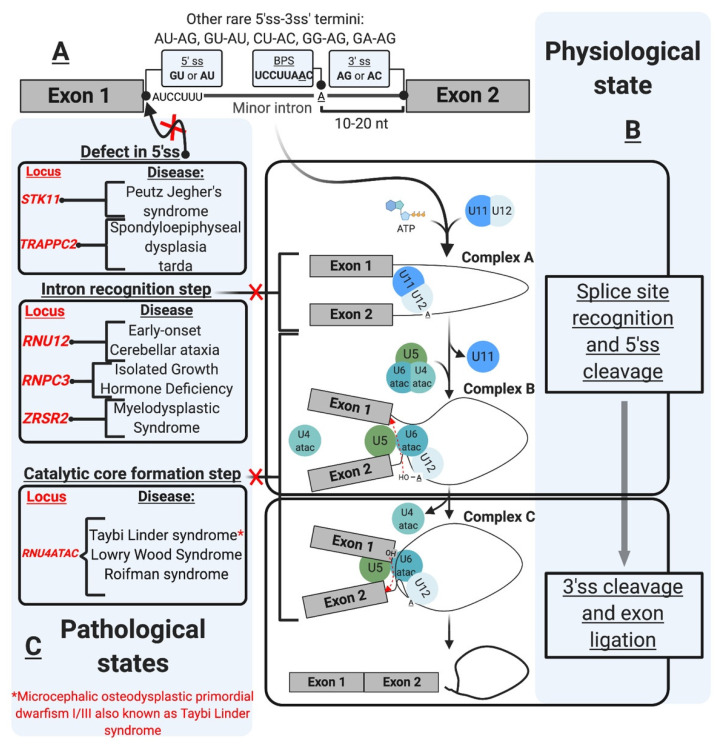
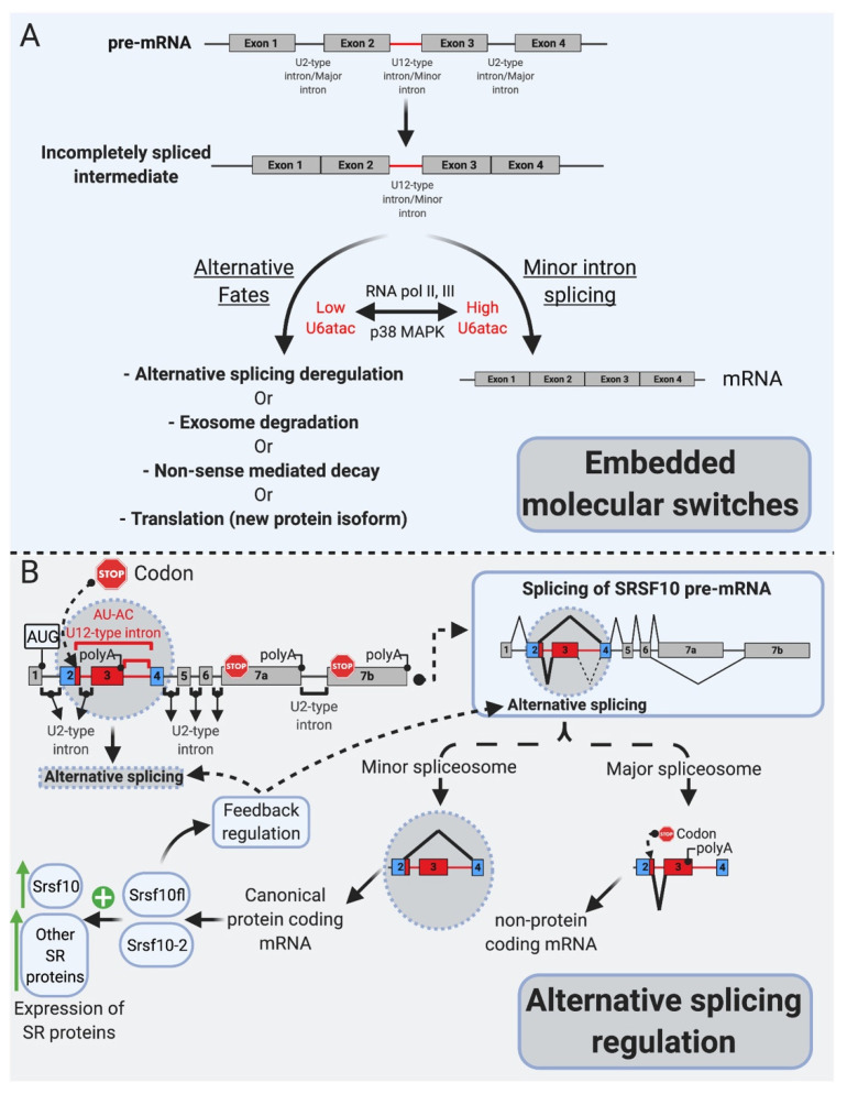
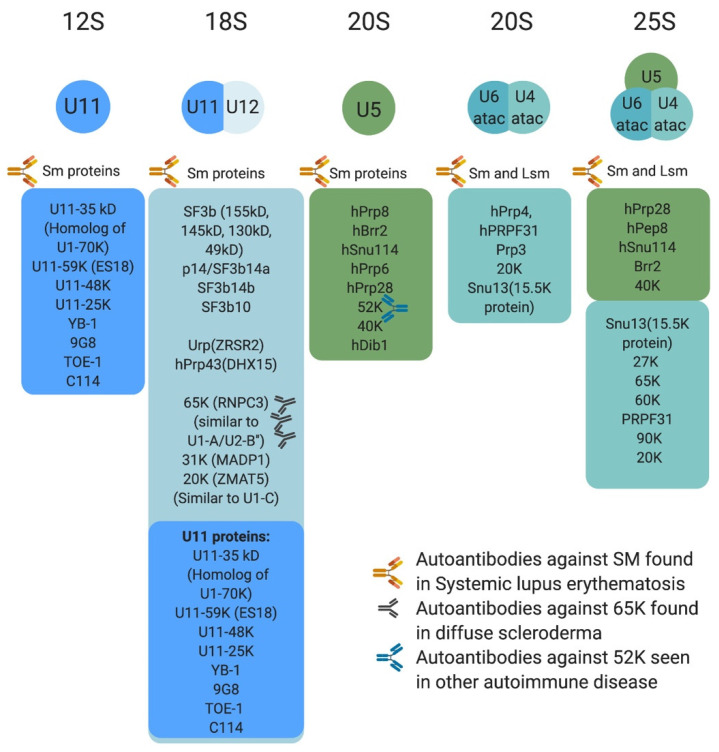
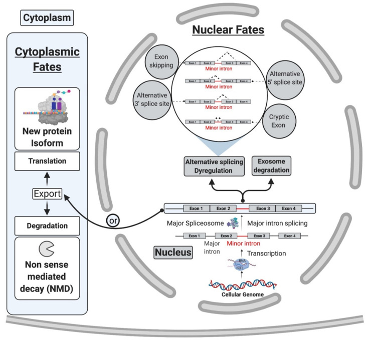
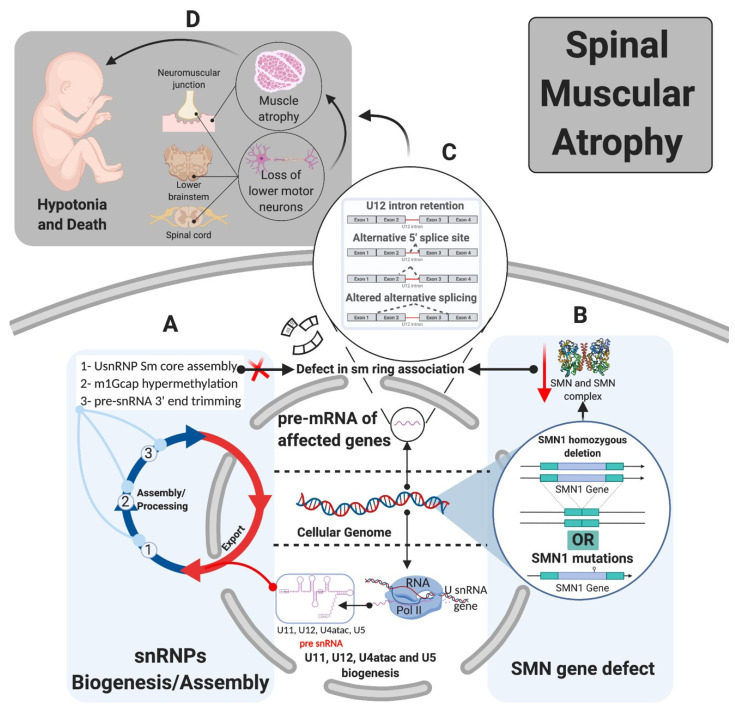
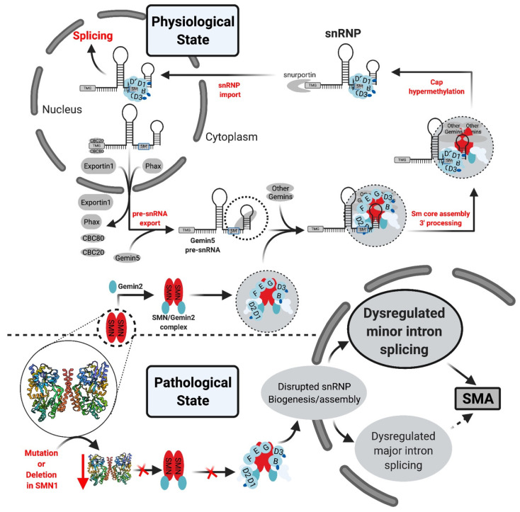
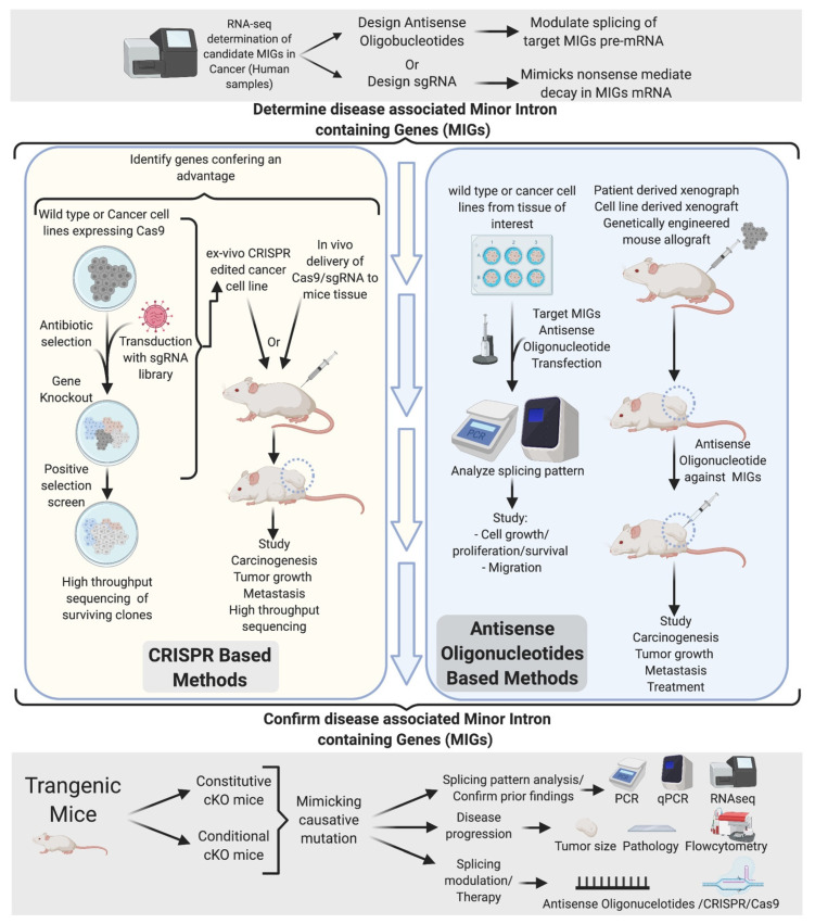
Pre-mRNA splicing is an essential step in gene expression and is catalyzed by two machineries in eukaryotes: the major (U2 type) and minor (U12 type) spliceosomes. While the majority of introns in humans are U2 type, less than 0.4% are U12 type, also known as minor introns (mi-INTs), and require a specialized spliceosome composed of U11, U12, U4atac, U5, and U6atac snRNPs. The high evolutionary conservation and apparent splicing inefficiency of U12 introns have set them apart from their major counterparts and led to speculations on the purpose for their existence. However, recent studies challenged the simple concept of mi-INTs splicing inefficiency due to low abundance of their spliceosome and confirmed their regulatory role in alternative splicing, significantly impacting the expression of their host genes. Additionally, a growing list of minor spliceosome-associated diseases with tissue-specific pathologies affirmed the importance of minor splicing as a key regulatory pathway, which when deregulated could lead to tissue-specific pathologies due to specific alterations in the expression of some minor-intron-containing genes. Consequently, uncovering how mi-INTs splicing is regulated in a tissue-specific manner would allow for better understanding of disease pathogenesis and pave the way for novel therapies, which we highlight in this review.

Keywords: RNA splicing; U12 introns; U2 introns; disease; minor introns; minor spliceosome.


Conflict of interest statement

The authors declare no conflict of interest.

Figures

Figure 1
Figure 1
The minor intron splicing pathway. (A). U12-type intron sequence characteristics [1,2,3,4,5,6]. These sequences were initially described in the literature based on DNA nomenclature. These were changed in the figure as the transcript depicted is an RNA transcript. The commonly observed 3′ and 5′ sequences of minor introns are shown in the boxes. The conserved sequences are shown at the 5′ ss and branchpoint (B). Spliceosomal assembly and the splicing reaction [7,8,9,10,11,12] occur as a two-step reaction. The interaction of the preformed U11/U12 di-snRNP leads to the formation of Complex A. U4atac/U6atac.U5 tri-snRNP allows the formation of Complex B, which carries the first transesterification reaction after the release of U4atac and U11. This reaction leads to the formation of Complex C, which carries the second transesterification reaction producing ligates exons and a minor intron lariat. (C). U12-type intron-associated diseases classification [13,14,15,16,17,18,19,20,21]. The diseases are depicted in association with the affected step. Diseases not shown in the figure include amyotrophic lateral sclerosis associated with mutations in fused in sarcoma (FUS) RNA-binding protein [22], and Noonan syndrome due to U12-type intron retention in LZTR1, a regulator of RAS-related GTPases [23].
Figure 2
Figure 2
mi-INTs physiologic roles. (A). mi-INTs function as embedded molecular switches of gene expression [25]. Instability of U6atac result in low U6atac levels, which leads to pre-mRNA transcripts with a retained mi-INT whose fates are depicted in Figure 3. Decreased transcription lowers U6atac levels and the expression of minor-intron-containing genes, while activated p38MAPK stabilizes U6atac, increasing its levels and allowing the proper splicing of mi-INTs, producing a full-length mRNA. (B). mi-INTs regulate alternative splicing through Srsf10 [24]. Low minor spliceosome activity causes an increase in the expression of non-protein-coding Srsf10 transcript through exon 3 inclusion as a consequence of major intron splicing. High minor spliceosome activity allows exclusion of exon 3, through splicing to exon 4, allowing formation of a protein-coding transcript. The functional Srsf10 autoregulates its expression and that of other SR proteins which regulate alternative splicing of thousands of downstream targets.
Figure 3
Figure 3
Protein composition of minor snRNPs and autoimmune conditions associated with them. Sm proteins include B/B’, D3, D2, D1, E, F, and G, whereas LSm proteins are LSm 2–8. The U4atac/U6atac.U5 tri-snRNP has two sets of Sm protein and one set of LSm proteins. Antibodies against some protein components of the minor spliceosome are found in systemic lupus erythematosus, diffuse scleroderma, and other autoimmune conditions.
Figure 4
Figure 4
Cytoplasmic and nuclear fates of pre-mRNA transcripts with retained mi-INTs [20,26,27]. mi-INTs retention may lead to trapping of the transcript within the nucleus. The trapped transcript may undergo exosomal degradation, activation of cryptic U2-type intron cryptic sites or exon skipping. Transcripts that escape degradation in the nucleus are exported to the cytoplasm they may be degraded by NMD or translated to a truncated or novel protein isoform.
Figure 5
Figure 5
Tissue specificity of mi-INTs-related diseases (SMA). (A). snRNP biogenesis and assembly requires SMN along with other proteins (SMN complex) for Sm core assembly, m1Gcap hypermethylation, and pre-snRNA 3’end trimming. (B). Loss or mutation in SMN1 with retention of SMN2 product allows the production of SMN at lower levels leading to lower SMN complex level and lower ability to interact with Sm proteins, preventing the formation of the Sm ring and mature snRNPs (major and minor) which preferentially affects minor snRNPs [28,29,30] leading to splicing defects. (C). Splicing defect in U2 and U12-type intron splicing with bias toward U12-type intron retention [29,31] leads to aberrant mRNA transcripts that may be degraded or produce aberrant proteins. (D). Phenotype of SMA depends on the severity of the illness, which is caused by motor neuron degeneration in the brain stem, anterior horn of the spinal cord, and muscle atrophy. This leads to hypotonia and eventual death [32,33].
Figure 6
Figure 6
U snRNP biogenesis pathway. In the physiological state, once the U snRNA is transcribed and capped it associates with multiple factors which form the export complex and allow U snRNA export into the cytoplasm. Subsequently, in a process mediated by SMN complex and Gemin proteins, the Sm proteins interact with the U snRNA at the Sm site. The SMN complex dissociates, followed by 3′ processing/trimming and cap hypermethylation. The final product is imported into the nucleus. Further modifications occur on the U snRNA, and snRNP (U1, U2, U4, U5 or U11/U12, U4atac)-specific proteins associate with U snRNP Sm core [34].
Figure 7
Figure 7
Experimental determination of disease-causing/-associated MIGs. Initial determination of disease-related MIGs through RNA-seq generates a list of potential candidates. Targeting of these candidates through CRISPR and ASOs in vivo and vitro narrows down the gene targets to a few. The ultimate confirmation comes from a genetically engineered mouse, mimicking the disease mutation found in human. This allows confirmation of mi-INTs splicing defects observed and determination of their contribution to the disease.

References

    1. Kruger K., Grabowski P.J., Zaug A.J., Sands J., Gottschling D.E., Cech T.R. Self-splicing RNA: Autoexcision and autocyclization of the ribosomal RNA intervening sequence of tetrahymena. Cell. 1982;31:147–157. doi: 10.1016/0092-8674(82)90414-7. - DOI - PubMed
    1. Guerrier-Takada C., Gardiner K., Marsh T., Pace N., Altman S. The RNA moiety of ribonuclease P is the catalytic subunit of the enzyme. Cell. 1983;35:849–857. doi: 10.1016/0092-8674(83)90117-4. - DOI - PubMed
    1. Wolff J.A., Malone R.W., Williams P., Chong W., Acsadi G., Jani A., Felgner P.L. Direct gene transfer into mouse muscle in vivo. Science. 1990;247:1465–1468. doi: 10.1126/science.1690918. - DOI - PubMed
    1. Martinon F., Krishnan S., Lenzen G., Magné R., Gomard E., Guillet J.-G., Lévy J.-P., Meulien P. Induction of virus-specific cytotoxic T lymphocytesin vivo by liposome-entrapped mRNA. Eur. J. Immunol. 1993;23:1719–1722. doi: 10.1002/eji.1830230749. - DOI - PubMed
    1. Polack F.P., Thomas S.J., Kitchin N., Absalon J., Gurtman A., Lockhart S., Perez J.L., Marc G.P., Moreira E.D., Zerbini C., et al. Safety and Efficacy of the BNT162b2 mRNA Covid-19 Vaccine. N. Engl. J. Med. 2020;383:2603–2615. doi: 10.1056/NEJMoa2034577. - DOI - PMC - PubMed

LinkOut - more resources