. 2021 Jun 4;22(11):6073.
doi: 10.3390/ijms22116073.

Exploring the Physiological Role of Transthyretin in Glucose Metabolism in the Liver

Affiliations

Exploring the Physiological Role of Transthyretin in Glucose Metabolism in the Liver

Mobina Alemi et al. Int J Mol Sci. .

Abstract

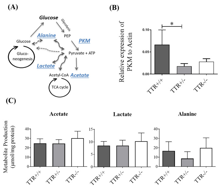
Transthyretin (TTR), a 55 kDa evolutionarily conserved protein, presents altered levels in several conditions, including malnutrition, inflammation, diabetes, and Alzheimer's Disease. It has been shown that TTR is involved in several functions, such as insulin release from pancreatic β-cells, recovery of blood glucose and glucagon levels of the islets of Langerhans, food intake, and body weight. Here, the role of TTR in hepatic glucose metabolism was explored by studying the levels of glucose in mice with different TTR genetic backgrounds, namely with two copies of the TTR gene, TTR+/+; with only one copy, TTR+/-; and without TTR, TTR-/-. Results showed that TTR haploinsufficiency (TTR+/-) leads to higher glucose in both plasma and in primary hepatocyte culture media and lower expression of the influx glucose transporters, GLUT1, GLUT3, and GLUT4. Further, we showed that TTR haploinsufficiency decreases pyruvate kinase M type (PKM) levels in mice livers, by qRT-PCR, but it does not affect the hepatic production of the studied metabolites, as determined by 1H NMR. Finally, we demonstrated that TTR increases mitochondrial density in HepG2 cells and that TTR insufficiency triggers a higher degree of oxidative phosphorylation in the liver. Altogether, these results indicate that TTR contributes to the homeostasis of glucose by regulating the levels of glucose transporters and PKM enzyme and by protecting against mitochondrial oxidative stress.

Keywords: glucose metabolism; glucose transporters; liver; mitochondria; transthyretin.


Conflict of interest statement

The authors declare no conflict of interest.

Figures

Figure 1
Figure 1
Effect of TTR and glucose on each other. (A) In mice, plasma glucose levels were shown to be higher in TTR+/− mice (N = 11), while TTR−/− (N = 7) showed no alteration as compared to plasmas from TTR+/+ animals (N = 5). (B) In primary hepatocytes, glucose levels, normalized by mg protein, were also higher in the extracellular culture media of primary hepatocytes derived from TTR+/− and TTR−/− mice. Further addition of h rTTR to the media of TTR+/− hepatocytes resulted in decreased glucose levels, while it did not alter glucose concentration in hepatocytes from TTR−/− mice. (C) In primary hepatocytes, different concentrations of glucose did not produce any alteration in the concentration of secreted TTR (N = 3 for each condition). Data are expressed as mean ± SD. Significant results are indicated by * when p < 0.05, ** when p < 0.01, *** when p < 0.001 and **** when p < 0.0001.
Figure 2
Figure 2
Effect of TTR on hepatic glucose transporters, assessed by qRT-PCR, Western blot, and immunocytochemistry. (A) In mice, at mRNA level, haploinsufficiency of TTR in TTR+/− mice livers significantly decreased expression of GLUT1, GLUT3, and GLUT4, compared to TTR+/+ livers, while TTR deficiency in TTR−/− mice livers showed lower expression of GLUT3 and GLUT4 and higher expression of GLUT2, compared to TTR+/+ livers (N = 4 for each genotype). (B) In primary hepatocytes, and at protein levels, cells derived from TTR+/− and TTR−/− mice showed lower expression of GLUT4 (N = 3 for each genotype). (C) HepG2 cells also showed higher protein levels of GLUT1 when incubated with h rTTR (0.2 µM), compared to untreated cells (N = 4 for each condition). Data are expressed as mean ± SD. Significant results are indicated by * when p < 0.05, ** when p < 0.01, and *** when p < 0.001.
Figure 3
Figure 3
Effect of TTR on the expression of liver PKM enzyme and hepatic production of glucose-derived metabolites. (A) A simple schematic representation of the metabolic pathways of glucose: during glycolysis, glucose is converted to pyruvate, through the action of PKM on PEP, producing ATP. Pyruvate can further turn into acetate, lactate, and alanine and/or enter the mitochondrial TCA cycle for further production of energy. Glucose can also be produced through gluconeogenesis in the liver. (B) In mouse livers, the relative expression of PKM, assessed by qRT-PCR, was shown to be lower in TTR+/− livers compared to those from TTR+/+ animals (N = 4 for each genotype). (C) In primary hepatocytes, the production of acetate, lactate, and alanine was assessed by 1H-NMR spectroscopy. No significant differences were observed in their production by hepatocytes from different TTR backgrounds mice (N = 6 for each condition). Data are expressed as mean ± SD. Significant results are indicated by * when p < 0.05.
Figure 4
Figure 4
Effect of TTR on mitochondria as assessed by fluorescent microscopy and Western blot. (A) HepG2 cells treated with h rTTR showed significantly higher mitochondrial density compared to untreated ones (N = 5 for each condition). (B) In mouse liver, and as assessed by Western blot, higher protein levels of mitochondrial CI and CII were observed in TTR+/− mice, and higher CII and CIII in TTR−/− mice, compared to TTR+/+ animals (N = 3 for each genotype). Data are expressed as mean ± SD. Significant results are indicated by * when p < 0.05, ** when p < 0.01, and *** when p < 0.001.
Figure 5
Figure 5
Graphical summary of the results obtained in the present study, illustrating the effects of TTR insufficiency on glucose metabolism of the liver. Insufficiency of TTR, which can result from its tetrameric instability and consequent increased clearance, can increase extra-hepatic glucose levels (1), which can be, at least partially, due to its negative effect on the expression of influx glucose transporters, GLUT1, GLUT3, and GLUT4 (2) and increased expression of efflux glucose transporter, GLUT2 (3). Moreover, the decrease in the expression of glycolytic enzyme, PKM (4), and the increase in mitochondrial CI and CII of the electron transport chain (5) are observed in the TTR haploinsufficiency mouse model, which in turn leads to higher levels of ROS and/or lipid peroxidation and ends up compromising the mitochondrial density and function (6).

References

    1. Taub R. Liver Regeneration: From Myth to Mechanism. Nat. Rev. Mol. Cell Biol. 2004;5:836–847. doi: 10.1038/nrm1489. - DOI - PubMed
    1. Rui L. Energy Metabolism in the Liver. Compr. Physiol. 2014;4:177–197. doi: 10.1002/cphy.c130024. - DOI - PMC - PubMed
    1. Grimm A., Friedland K., Eckert A. Mitochondrial Dysfunction: The Missing Link between Aging and Sporadic Alzheimer’s Disease. Biogerontology. 2015 doi: 10.1007/s10522-015-9618-4. - DOI - PubMed
    1. Saxena A.K., Srivastava P., Kale R.K., Baquer N.Z. Impaired Antioxidant Status in Diabetic Rat Liver. Effect of Vanadate. Biochem. Pharmacol. 1993;45:539–542. doi: 10.1016/0006-2952(93)90124-F. - DOI - PubMed
    1. Guo C., Sun L., Chen X., Zhang D. Oxidative Stress, Mitochondrial Damage and Neurodegenerative Diseases. Neural Regen. Res. 2013;8:2003–2014. doi: 10.3969/j.issn.1673-5374.2013.21.009. - DOI - PMC - PubMed