. 2021 Jun 4;10(6):703.
doi: 10.3390/pathogens10060703.

Cystic Fibrosis Sputum Impairs the Ability of Neutrophils to Kill Staphylococcus aureus

Affiliations

Cystic Fibrosis Sputum Impairs the Ability of Neutrophils to Kill Staphylococcus aureus

Kayla Fantone et al. Pathogens. .

Abstract

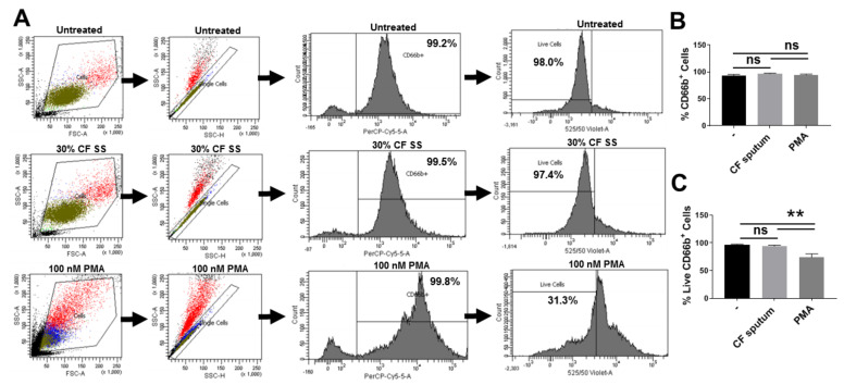
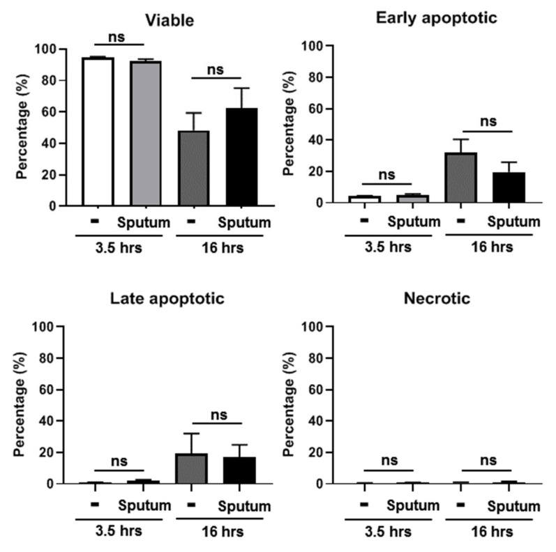
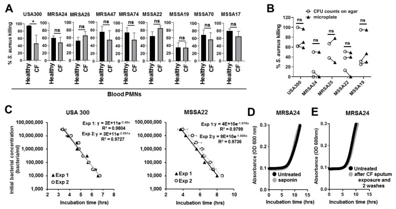
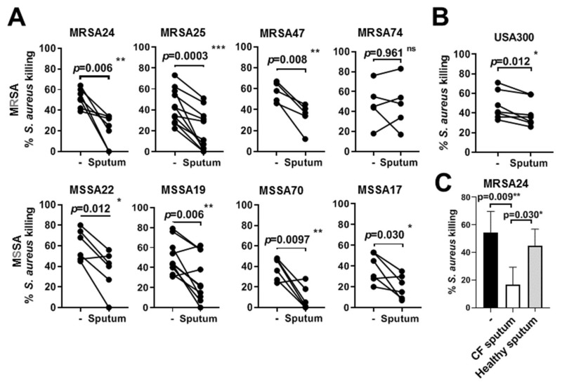
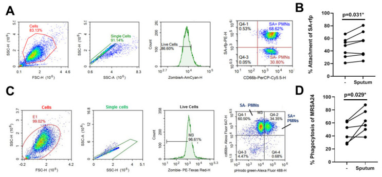
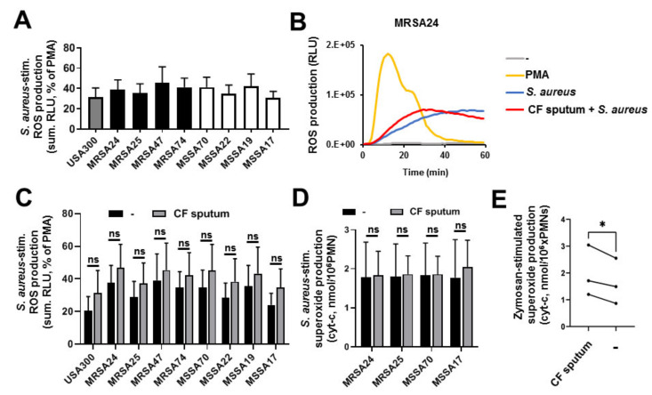
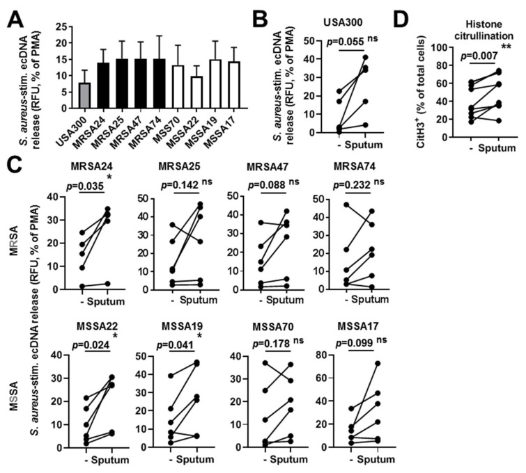
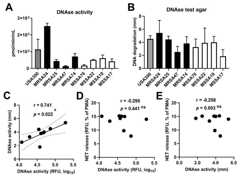
Cystic fibrosis (CF) airway disease is characterized by chronic microbial infections and infiltration of inflammatory polymorphonuclear (PMN) granulocytes. Staphylococcus aureus (S. aureus) is a major lung pathogen in CF that persists despite the presence of PMNs and has been associated with CF lung function decline. While PMNs represent the main mechanism of the immune system to kill S. aureus, it remains largely unknown why PMNs fail to eliminate S. aureus in CF. The goal of this study was to observe how the CF airway environment affects S. aureus killing by PMNs. PMNs were isolated from the blood of healthy volunteers and CF patients. Clinical isolates of S. aureus were obtained from the airways of CF patients. The results show that PMNs from healthy volunteers were able to kill all CF isolates and laboratory strains of S. aureus tested in vitro. The extent of killing varied among strains. When PMNs were pretreated with supernatants of CF sputum, S. aureus killing was significantly inhibited suggesting that the CF airway environment compromises PMN antibacterial functions. CF blood PMNs were capable of killing S. aureus. Although bacterial killing was inhibited with CF sputum, PMN binding and phagocytosis of S. aureus was not diminished. The S. aureus-induced respiratory burst and neutrophil extracellular trap release from PMNs also remained uninhibited by CF sputum. In summary, our data demonstrate that the CF airway environment limits killing of S. aureus by PMNs and provides a new in vitro experimental model to study this phenomenon and its mechanism.

Keywords: PMN; Staphylococcus aureus; cystic fibrosis; killing; neutrophil extracellular traps; respiratory burst; sputum.


Conflict of interest statement

The authors have no conflict of interest related to this work.

Figures

Figure 1
Figure 1
CF sputum supernatant treatment does not affect PMN viability. PMN purity and viability following isolation from blood and subsequent 3.5 h-CF sputum incubation was measured by flow cytometry using the Zombie Aqua Cell Viability Kit™. (A) Representative images of the gating strategy used to determine the percent of viable PMNs (CD66b+/Zombie Aqua) for each condition tested are shown. (B) Flow cytometric analysis showed no significant difference in the percent of PMNs and CD66 surface expression among the conditions tested (n = 15). (C) CF sputum treatment does not impair PMN viability under the conditions used in this study. Treatment with 100 nM PMA for 30 min, however, results in a significant decrease in PMN viability compared to both the sputum supernatant-treated and untreated PMNs (n = 15). One-way ANOVA, Tukey’s multiple comparison test. **, p < 0.01; ns, not significant. PMA, phorbol 12-myristate 13-acetate; CF SS; Cystic fibrosis sputum supernatant.
Figure 2
Figure 2
CF sputum supernatant does not induce apoptosis or necrosis in human PMNs. Human blood PMNs were incubated in CF sputum supernatants for 3.5 or 16 h prior to fluorescence staining with Apotracker (TM) Green apoptosis probe and propidium iodide (PI) viability dye. The following cell populations were identified by flow cytometry: viable (double negative), early apoptotic (PI-negative, Apotracker-positive), necrotic (PI-positive, Apotracker-negative) and late apoptotic (double positive). Results are expressed as mean ± S.E.M (n = 6). One-way ANOVA, Dunn’s multiple comparison test. ns, not significant.
Figure 3
Figure 3
Healthy and CF human blood neutrophils kill CF isolates of S. aureus in vitro. (A) Healthy and CF human blood PMNs were infected with the S. aureus reference strain (USA300), 4 MRSA isolates, and 4 MSSA isolates collected from CF patients. All isolates were infected at MOI of 10 except MSSA70 and MSSA17 that were infected at MOI of 5. Bacterial killing was measured by the high-throughput microplate-based assay. Mean ± S.E.M, n = 6. Two-tailed, paired Students’ t-test. (B) Comparison of PMN-mediated S. aureus killing by two methods: high-throughput microplate-based assay and agar plate based colony counting assay. Lines connect data derived from the same, individual experiment and same samples via the two different methods. Experiments were repeated three times on three independent human donors’ neutrophils. (C) Standard curves of USA 300 and MSSA22 growth calibrations are shown as examples indicating the tight correlation between initial bacterial concentrations (y axis) and the incubation time values (x axis). Two representative curves for each strain are shown in the same graph with trend lines, the equation and R2 values. Mean ± S.D. (D) Representative growth curves of MRSA24 with or without saponin treatment that is used in the killing assay (n = 3). (E) Representative growth curves of MRSA24 without any treatment or in medium that was used to resuspend human PMNs after CF sputum exposure for 3.5 h and two washes according to the protocol of the killing assay. These data show that two washes of human PMNS are sufficient to ensure that the CF sputum exposure of PMNs does not interfere with subsequent growth of tested bacteria. CFU, colony-forming unit; ns, not significant; OD, optical density.
Figure 4
Figure 4
CF sputum treatment impairs neutrophils’ ability to kill S. aureus. PMNs were first treated with 30% (v/v) CF sputum cocktail and then infected with the S. aureus lab strain (USA300), 4 MRSA isolates and 4 MSSA isolates collected from CF patients. Bacterial killing was measured by a high-throughput microplate-based assay. (A) The effect of CF sputum pretreatment on PMN-mediated killing of S. aureus CF clinical isolates (n = 5–11) or (B) USA 300 (n = 7) is shown. All isolates were infected at MOI of 10 except MSSA70 and MSSA17 were infected at MOI of 5. Mean ± S.E.M. Data were analyzed by Wilcoxon matched-pairs signed rank test. (C) The effect of pretreatment with sputum cocktails isolated and pooled from CF patients or healthy controls (n = 2) on PMN-mediated killing of MRSA24 (n = 4). Mean ± S.E.M. Two-tailed, paired Students’ t-test (A,B) or one-way ANOVA test (C) were used. Statistically significant differences were considered as *, p < 0.05; **, p < 0.01; ***, p < 0.001. Ns, not significant.
Figure 5
Figure 5
CF sputum treatment does not impair S. aureus binding and phagocytosis by PMNs. PMNs were isolated from healthy donors and exposed to CF sputum supernatant. To measure bacterial attachment, PMNs were infected with fluorescently labeled, opsonized S. aureus (SA-rfp) (10 MOI). (A) Representative images of the gating strategy used to determine the percent of SA-rfp attached PMNs (CD66b+/Zombie Aqua-) for each condition. (B) Comparison of the attachment of SA-rfp to PMNs untreated or treated with CF sputum supernatant (n = 7). (C) To determine phagocytosis, MRSA24 CF isolate was labelled with the pH-sensitive dye pHrodo, opsonized and exposed to PMNs. Representative images of the gating strategy used to determine the percent of MRSA24 phagocytosed by PMNs for each condition. (D) Comparison of MRSA24 phagocytosis by PMNs that were untreated or treated with CF sputum supernatant (n = 6). Two-tailed, paired Students’ t-test. *, p < 0.05.
Figure 6
Figure 6
CF sputum does not impair PMN superoxide production in response to CF isolates of S. aureus. Human blood PMNs were exposed to the indicated isolates of S. aureus (10 MOI) and ROS production was measured by Diogenes-based chemiluminescence (AC) or cytochrome-c reduction assay (DE). (A) PMNs respond to the presence of CF bacterial isolates with robust ROS production (mean ± S.E.M, n = 6–7). (B) Representative kinetics of S. aureus-stimulated PMN ROS release curves (60 min) (n = 7). In total, 100 nM PMA was used as a positive control. The effect of CF sputum exposure on (C) ROS production or (D) extracellular superoxide generation in human PMNs exposed to the indicated isolates of S. aureus is shown. ROS production was calculated for 60 min (n = 6–7) by Diogenes-based chemiluminescence while superoxide production was measured for 60 min by the cytochrome-c reduction assay (n = 3). Mean ± S.E.M. Data were analyzed by Wilcoxon matched-pairs signed rank test. (E) Comparison of superoxide production by sputum-treated PMNs vs. sputum-untreated cells following exposure to zymosan (10 MOI, opsonized). Two-tailed, paired Students’ t-test. Statistically significant differences were considered as *, p < 0.05. Ns, not significant.
Figure 7
Figure 7
NET formation triggered by CF isolates of S. aureus is not compromised by CF sputum. Human blood PMNs were exposed to the indicated isolates of S. aureus (10 MOI) and extracellular DNA (ecDNA) release was measured for up to 8 h in presence of the membrane-impermeable, DNA-sensitive fluorescent dye, Sytox Orange. (A) EcDNA release in S. aureus-stimulated PMNs after 8 h measured as increase in fluorescence. The signal by unstimulated PMNs was subtracted and the S. aureus-induced ecDNA signal was normalized on the signal obtained by PMA stimulation (100 nM). Mean ± S.E.M, n = 5–6. (B) The effect of CF sputum treatment on ecDNA release in PMNs exposed to USA300. Mean ± S.E.M, n = 6. (C) The effect of CF sputum treatment on ecDNA release in PMNs exposed to the indicated CF isolates of S. aureus. Mean ± S.E.M, n = 6. (D) Histone H3 citrullination in PMNs exposed to the CF sputum in the absence of bacterial stimulation measured by flow cytometry (n = 9). Two-tailed, paired Students’ t-test. Statistically significant differences were considered as *, p < 0.05; **, p < 0.01. Ns, not significant.
Figure 8
Figure 8
DNAse activity of CF clinical isolates of S. aureus. DNAse activity was measured in the indicated CF isolates of S. aureus by (A) a fluorescent enzymatic activity assay (n = 3–6), or (B) a DNAse I agar plate-based assay (n = 3). Mean ± S.E.M. Correlation analysis is shown between (C) the two DNAse activity measures, (D) NET release and results of the fluorescent DNAse activity assay, and ® NET release and results of the plate-based DNAse activity assay. Pearson’s correlation coefficient(r). Statistically significant differences were considered as *, p < 0.05. Ns, not significant.

Similar articles

Cited by

References

    1. Ahlgren H.G., Benedetti A., Landry J.S., Bernier J., Matouk E., Radzioch D., Lands L.C., Rousseau S., Nguyen D. Clinical outcomes associated with Staphylococcus aureus and Pseudomonas aeruginosa airway infections in adult cystic fibrosis patients. BMC Pulm. Med. 2015;15:67. doi: 10.1186/s12890-015-0062-7. - DOI - PMC - PubMed
    1. Cystic Fibrosis Foundation Patient Registry . 2017 Annual Data Report. Cystic Fibrosis Foundation Patient Registry; Bethesda, MD, USA: 2018.
    1. Akil N., Muhlebach M.S. Biology and management of methicillin resistant Staphylococcus aureusin cystic fibrosis. Pediatr. Pulmonol. 2018;53:S64–S74. doi: 10.1002/ppul.24139. - DOI - PubMed
    1. Lo D.K., Muhlebach M.S., Smyth A.R. Interventions for the eradication of meticillin-resistant Staphylococcus aureus (MRSA) in people with cystic fibrosis. Cochrane Database Syst. Rev. 2018;7:CD009650. doi: 10.1002/14651858.CD009650.pub4. - DOI - PMC - PubMed
    1. Muhlebach M.S., Zorn B.T., Esther C.R., Hatch J.E., Murray C.P., Turkovic L., Ranganathan S.C., Boucher R.C., Stick S.M., Wolfgang M.C. Initial acquisition and succession of the cystic fibrosis lung microbiome is associated with disease progression in infants and preschool children. PLoS Pathog. 2018;14:e1006798. doi: 10.1371/journal.ppat.1006798. - DOI - PMC - PubMed

LinkOut - more resources