. 2021 Jun 4;9(6):643.
doi: 10.3390/biomedicines9060643.

Palmitoleate Protects against Zika Virus-Induced Placental Trophoblast Apoptosis

Affiliations

Palmitoleate Protects against Zika Virus-Induced Placental Trophoblast Apoptosis

Philma Glora Muthuraj et al. Biomedicines. .

Abstract

Zika virus (ZIKV) infection in pregnancy is associated with the development of microcephaly, intrauterine growth restriction, and ocular damage in the fetus. ZIKV infection of the placenta plays a crucial role in the vertical transmission from the maternal circulation to the fetus. Our previous study suggested that ZIKV induces endoplasmic reticulum (ER) stress and apoptosis of placental trophoblasts. Here, we showed that palmitoleate, an omega-7 monounsaturated fatty acid, prevents ZIKV-induced ER stress and apoptosis in placental trophoblasts. Human trophoblast cell lines (JEG-3 and JAR) and normal immortalized trophoblasts (HTR-8) were used. We observed that ZIKV infection of the trophoblasts resulted in apoptosis and treatment of palmitoleate to ZIKV-infected cells significantly prevented apoptosis. However, palmitate (saturated fatty acid) did not offer protection from ZIKV-induced ER stress and apoptosis. We also observed that the Zika viral RNA copies were decreased, and the cell viability improved in ZIKV-infected cells treated with palmitoleate as compared to the infected cells without palmitoleate treatment. Further, palmitoleate was shown to protect against ZIKV-induced upregulation of ER stress markers, C/EBP homologous protein and X-box binding protein-1 splicing in placental trophoblasts. In conclusion, our studies suggest that palmitoleate protects placental trophoblasts against ZIKV-induced ER stress and apoptosis.

Keywords: Zika virus; apoptosis; endoplasmic reticulum stress; monounsaturated fatty acids; placenta; trophoblast; viral replication.


Conflict of interest statement

The authors declare no conflict of interest.

Figures

Figure 1
Figure 1
Palmitoleate treatment after Zika virus (ZIKV) PRVABC59 strain (PRV) infection inhibits apoptosis in JEG-3 cells. (A) Structural and biochemical characterization of apoptosis in JEG-3 infected with the 1.0 MOI (multiplicity of infection) ZIKV PRV strain, 48–72 h showed an increase in percent apoptotic nuclei compared to uninfected vehicle cells. Infected cells treated with 100 or 200 µM of palmitoleate, 48–72 hpi (hours post infection) showed a decrease in the percent apoptotic nuclei when compared to ZIKV-infected cells alone (left panels). (B) There was a significant activation of caspase 3/7 in ZIKV-infected cells, but caspase 3/7 activation was significantly blocked when the infected cells were treated with 100 or 200 µM of palmitoleate both at 48 and 72 hpi (right panels). Data represent mean ± SEM, n = 3. * p < 0.05 compared to uninfected vehicles cells, # p < 0.05 compared to ZIKV-infected cells.
Figure 2
Figure 2
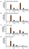
Treatment of the placental cells with palmitoleate after MR766 (MRV) strain of Zika virus (ZIKV) infection inhibits apoptosis. (A) Phase contrast (left panels) and DAPI stained (right panels) image panels: characteristic nuclear morphological changes were detected with DAPI staining in third trimester-derived JAR cells after 48 hours post-infection (hpi) with MRV. Vehicle cells show little to no DAPI staining. Infected panel shows higher number of DAPI stained fragmented and condensed nuclei compared to 100 or 200 µM palmitoleate treated cells. The images shown here are representative images. (B) JAR cells infected with 1.0 MOI MRV strain of ZIKV showed increase in the percentage apoptotic nuclei when compared to uninfected vehicle cells. In cells treated with 100 and 200 µM palmitoleate 48 hpi showed a significant decrease in the percent apoptotic nuclei compared to ZIKV-infected cells. (C) There was a significant increase in caspase 3/7 activity in MRV-infected cells, but this was significantly blocked when the cells were treated with 200 µM of palmitoleate post-infection and a trend towards downregulation at 100 µM palmitoleate treatment in the MRV-infected cells. (D) Characteristic nuclear morphological changes were detected using DAPI staining in the first trimester derived HTR-8 cells after 72 hpi were calculated and represented as percent apoptotic nuclei. Cells infected with 1.0 MOI r-MRV (recombinant MR766) strain of ZIKV showed an increase in the percent apoptotic nuclei when compared to uninfected vehicle cells. However, cells treated with 100 and 200 µM of palmitoleate post-infection showed a significant decrease in the percentage apoptotic nuclei compared to ZIKV-infected cells. (E) There was an increase in caspase 3/7 activity in ZIKV infected cells when compared to uninfected vehicle cells, but this activation was significantly blocked when the cells were treated with 100 or 200 µM of palmitoleate, 72 hpi. Data represent mean ± SEM, n = 3 for percent apoptotic nuclei, and n = 4 for caspase 3/7 activity. * p < 0.05 compared to uninfected vehicles cells, # p < 0.05 compared to ZIKV-infected cells.
Figure 3
Figure 3
Treatment of the placental trophoblasts with palmitoleate after Zika virus (ZIKV) infection interferes with viral replication. The presence of the viral envelope RNA copy number in the cell culture supernatant of both (A) JAR and (B) HTR-8 cells infected with 0.1 and 1.0 MOI of ZIKV MR766 strain 72 h and 96 hpi, respectively. When MRV-infected cells were treated with palmitoleate (100–200 µM), there was significant reduction in the viral envelope RNA copy number in the cell culture supernatant with both 0.1 and 1 MOI in JAR cells and with 1 MOI in HTR-8 cells (A,B). The presence of viral envelope RNA copy number both in JAR (C) and HTR-8 (D) cell lysate infected with 0.1 and 1.0 MOI of ZIKV MR766 strain 72 and 96 hpi, respectively. When the infected cells were treated with 100–200 µM of palmitoleate, there was a trend showing reduced viral envelope RNA per microgram of total RNA in both JAR cell lysate (C) and HTR-8 cell lysates (D). Data represent mean ± SEM, n = 3. # p < 0.05 compared to ZIKV-infected cells. (EG) Viral E protein staining in JEG cells infected with 0.1 MOI r-MRV and in cells treated with 100 or 200µM palmitoleate, 48 hpi. (E) ZIKV-infected cells show green fluorescence indicating presence of viral E protein. (F) ZIKV-infected JEG-3 cells treated with 200 µM palmitoleate and show a reduction in the intensity of viral E protein staining. (G) Infected JEG-3 cells treated with 100–200 µM palmitate showed increased intensity of viral E protein staining. Nuclei were stained with DAPI. (H) Immunoblot analysis showed a dramatic increase in viral E protein expression with ZIKV infection in JEG-3 cells compared to vehicle uninfected cells. Treatment of 200 µM palmitoleate to ZIKV-infected cells showed reduced viral E protein expression compared to the ZIKV-infected cells alone.
Figure 3
Figure 3
Treatment of the placental trophoblasts with palmitoleate after Zika virus (ZIKV) infection interferes with viral replication. The presence of the viral envelope RNA copy number in the cell culture supernatant of both (A) JAR and (B) HTR-8 cells infected with 0.1 and 1.0 MOI of ZIKV MR766 strain 72 h and 96 hpi, respectively. When MRV-infected cells were treated with palmitoleate (100–200 µM), there was significant reduction in the viral envelope RNA copy number in the cell culture supernatant with both 0.1 and 1 MOI in JAR cells and with 1 MOI in HTR-8 cells (A,B). The presence of viral envelope RNA copy number both in JAR (C) and HTR-8 (D) cell lysate infected with 0.1 and 1.0 MOI of ZIKV MR766 strain 72 and 96 hpi, respectively. When the infected cells were treated with 100–200 µM of palmitoleate, there was a trend showing reduced viral envelope RNA per microgram of total RNA in both JAR cell lysate (C) and HTR-8 cell lysates (D). Data represent mean ± SEM, n = 3. # p < 0.05 compared to ZIKV-infected cells. (EG) Viral E protein staining in JEG cells infected with 0.1 MOI r-MRV and in cells treated with 100 or 200µM palmitoleate, 48 hpi. (E) ZIKV-infected cells show green fluorescence indicating presence of viral E protein. (F) ZIKV-infected JEG-3 cells treated with 200 µM palmitoleate and show a reduction in the intensity of viral E protein staining. (G) Infected JEG-3 cells treated with 100–200 µM palmitate showed increased intensity of viral E protein staining. Nuclei were stained with DAPI. (H) Immunoblot analysis showed a dramatic increase in viral E protein expression with ZIKV infection in JEG-3 cells compared to vehicle uninfected cells. Treatment of 200 µM palmitoleate to ZIKV-infected cells showed reduced viral E protein expression compared to the ZIKV-infected cells alone.
Figure 4
Figure 4
Palmitoleate protects against ZIKV-induced apoptosis but not palmitate. (A,B) There was significant reduction in (A) percentage apoptotic nuclei and (B) caspase 3/7 activation in JEG-3 cells treated with palmitoleate 48 hpi with 0.1 r-MRV suggesting the protective role of palmitoleate against ZIKV-induced apoptosis. When the JEG-3 cells were treated with palmitic acid 48 hpi, there was significant increase in the percent apoptotic nuclei and a trend towards increase in caspase 3/7 activation, suggesting that palmitate did not protect against ZIKV-induced apoptosis. Data represent mean ± SEM, n = 3 for percentage apoptotic nuclei and n = 4 for caspase 3/7 activity. * p < 0.05 compared to uninfected vehicles cells, # p < 0.05 compared to ZIKV-infected cells. (C) Similarly, in JEG-3 cells with 1.0 MOI of PRV for 48 h the percent apoptotic nuclei significantly reduced in 100–200 µM palmitoleate treated PRV-infected cells compared to PRV-infected cells alone; however, treatment of palmitate to ZIKV-infected cells did not prevent ZIKV-induced apoptosis. Data represent mean ± SEM, n = 3. * p < 0.05 compared to uninfected vehicles cells, # p < 0.05 compared to ZIKV-infected cells.
Figure 5
Figure 5
Palmitoleate improves cell viability in Zika virus (ZIKV)-infected cells. (A) The percent cell survival using crystal violet assay significantly reduced with 0.1MOI r-MRV when compared to uninfected vehicle cells. The percent cell survival significantly increased with 100 or 200 µM of palmitoleate treatment but was not seen with 100–200 µM palmitate treatment, 48 hpi. (B) The percent cell survivability using MTT showed significant improvement in cell survivability with 200 µM palmitoleate treatment in ZIKV-infected cells. Whereas 200 µM palmitate treatment in ZIKV-infected cells also showed a significant increase in cell survivability. Data represent mean ± SEM, n = 3 for percent cell viability using crystal violet assay and n = 4 for MTT assay. * p < 0.05 compared to uninfected vehicles cells, # p < 0.05 compared to ZIKV-infected cells.
Figure 6
Figure 6
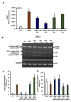
Palmitoleate protects against ZIKV-induced ER stress but not palmitate. (A) CHOP mRNA expression was significantly upregulated with 0.1 MOI r-MRV 48 hpi in JEG-3 cells compared to uninfected control cells. There was significant reduction in CHOP mRNA expression in JEG-3 cells treated with 200 µM of palmitoleate 48hpi with ZIKV suggesting protective role of palmitoleate against ZIKV-induced apoptosis and a trend towards decreased expression with 100 µM palmitoleate treatment, but this was not seen with the treatment of 100 or 200 µM palmitate to r-MRV-infected cells. (B) Similarly, there was increase in XBP1 mRNA splicing in infected cells (band ~448 bp) when compared to uninfected vehicle cells. There was significant inhibition of XBP1 mRNA splicing with the treatment of 100 or 200 µM of palmitoleate in ZIKV-infected cells. However, the protective properties were not seen with the treatment of palmitate to ZIKV-infected cells. (C) Quantified levels of spliced XBP1 mRNA (448 bp) relative to GAPDH (Spliced XPB1/GAPDH). (D) Quantified levels of unspliced XBP1 mRNA (474 bp) relative to GAPDH (Unspliced XPB1/GAPDH). Data represent mean ± SEM, n = 3. * p < 0.05 compared to uninfected vehicles cells, # p < 0.05 compared to ZIKV-infected cells.
Figure 7
Figure 7
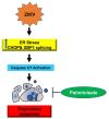
The schematic diagram represents palmitoleate protection against Zika virus (ZIKV)-induced ER stress and apoptosis in placental trophoblasts. ZIKV infection in trophoblasts elicits ER stress via the upregulation of CHOP and XBP1 mRNA splicing, which in turn activates apoptosis. Supplementation of palmitoleate protects against ZIKV-induced ER stress and trophoblast apoptosis.

Similar articles

Cited by

References

    1. Dick G.W.A., Kitchen S.F., Haddow A.J. Zika Virus (I). Isolations and serological specificity. Trans. R. Soc. Trop. Med. Hyg. 1952;46:509–520. doi: 10.1016/0035-9203(52)90042-4. - DOI - PubMed
    1. Weaver S.C., Costa F., Garcia-Blanco M.A., Ko A., Ribeiro G.S., Saade G., Shi P.-Y., Vasilakis N. Zika virus: History, emergence, biology, and prospects for control. Antivir. Res. 2016;130:69–80. doi: 10.1016/j.antiviral.2016.03.010. - DOI - PMC - PubMed
    1. Musso D., Gubler D.J. Zika Virus. Clin. Microbiol. Rev. 2016;29:487–524. doi: 10.1128/CMR.00072-15. - DOI - PMC - PubMed
    1. Moore C.A., Staples J.E., Dobyns W.B., Pessoa A., Ventura C.V., Da Fonseca E.B., Ribeiro E.M., Ventura L.O., Neto N.N., Arena J.F., et al. Characterizing the Pattern of Anomalies in Congenital Zika Syndrome for Pediatric Clinicians. JAMA Pediatr. 2017;171:288–295. doi: 10.1001/jamapediatrics.2016.3982. - DOI - PMC - PubMed
    1. Rosário M.S.D., De Siqueira I.C., Rodrigues S.G., Martins L.C., Vasilakis N., Novaes M.A.C., Alcantara L.C.J., Farias D.S., Jesus P.A., Ko A.I., et al. Guillain–Barré Syndrome After Zika Virus Infection in Brazil. Am. J. Trop. Med. Hyg. 2016;95:1157–1160. doi: 10.4269/ajtmh.16-0306. - DOI - PMC - PubMed