

Disruptions in Effective Connectivity within and between Default Mode Network and Anterior Forebrain Mesocircuit in Prolonged Disorders of Consciousness
- PMID: 34200092
- PMCID: PMC8227204
- DOI: 10.3390/brainsci11060749
Disruptions in Effective Connectivity within and between Default Mode Network and Anterior Forebrain Mesocircuit in Prolonged Disorders of Consciousness
Abstract
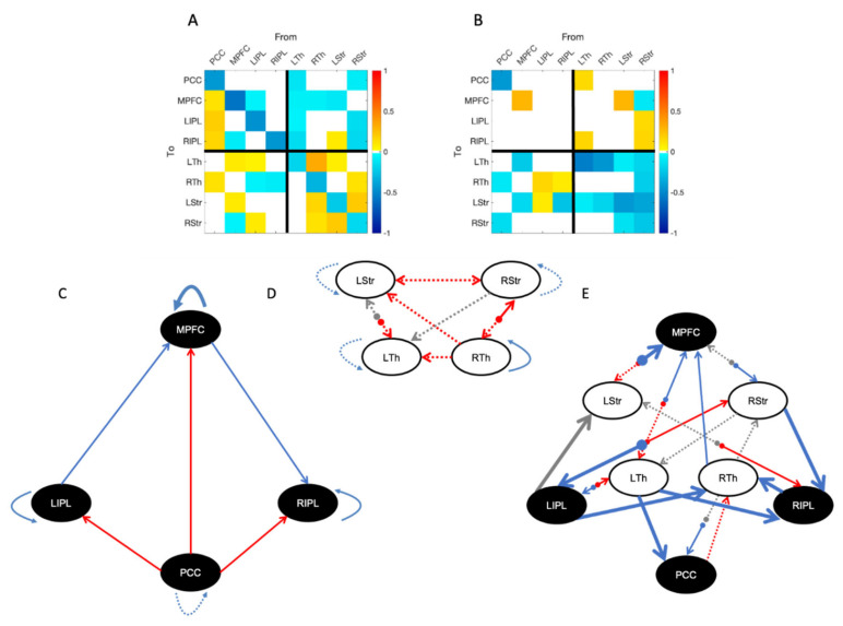
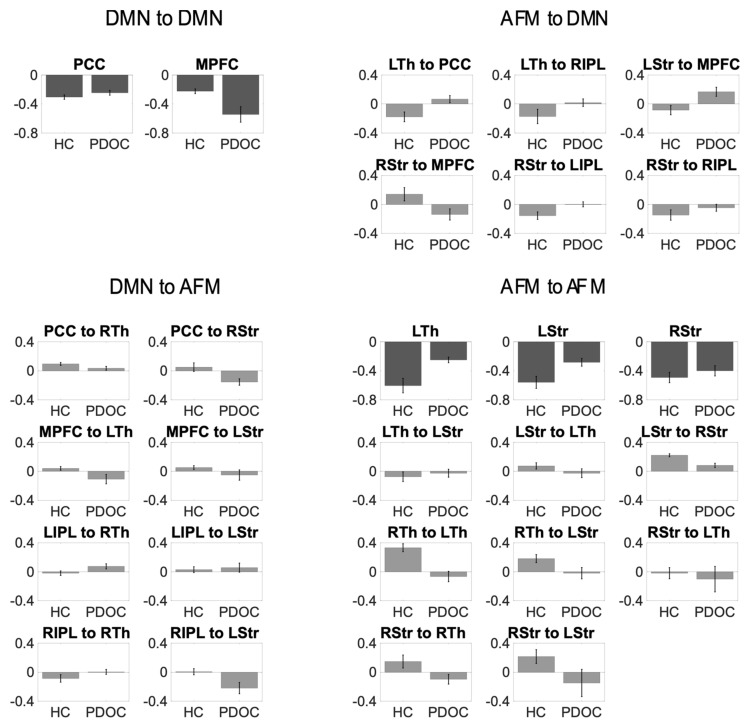
Recent research indicates prolonged disorders of consciousness (PDOC) result from structural and functional impairments to key cortical and subcortical networks, including the default mode network (DMN) and the anterior forebrain mesocircuit (AFM). However, the specific mechanisms which underpin such impairments remain unknown. It is known that disruptions in the striatal-pallidal pathway can result in the over inhibition of the thalamus and lack of excitation to the cortex that characterizes PDOC. Here, we used spectral dynamic causal modelling and parametric empirical Bayes on rs-fMRI data to assess whether DMN changes in PDOC are caused by disruptions in the AFM. PDOC patients displayed overall reduced coupling within the AFM, and specifically, decreased self-inhibition of the striatum, paired with reduced coupling from striatum to thalamus. This led to loss of inhibition from AFM to DMN, mostly driven by posterior areas including the precuneus and inferior parietal cortex. In turn, the DMN showed disruptions in self-inhibition of the precuneus and medial prefrontal cortex. Our results provide support for the anterior mesocircuit model at the subcortical level but highlight an inhibitory role for the AFM over the DMN, which is disrupted in PDOC.
Keywords: anterior forebrain mesocircuit; coma recovery scale-revised; default mode network; disorders of consciousness; dynamic causal modelling; effective connectivity; fMRI; minimally conscious state; parametric empirical Bayes; vegetative state.
Conflict of interest statement
The authors declare that the research was conducted in the absence of any commercial or financial relationships that could be construed as a potential conflict of interest.
Figures
Similar articles
-
Relationship between the anterior forebrain mesocircuit and the default mode network in the structural bases of disorders of consciousness.Neuroimage Clin. 2015 Nov 10;10:27-35. doi: 10.1016/j.nicl.2015.11.004. eCollection 2016. Neuroimage Clin. 2015. PMID: 26693399 Free PMC article.
-
Mapping the subcortical connectivity of the human default mode network.Neuroimage. 2021 Dec 15;245:118758. doi: 10.1016/j.neuroimage.2021.118758. Epub 2021 Nov 25. Neuroimage. 2021. PMID: 34838949 Free PMC article.
-
Exploration of resting-state brain functional connectivity as preclinical markers for arousal prediction in prolonged disorders of consciousness: A pilot study based on functional near-infrared spectroscopy.Brain Behav. 2024 Aug;14(8):e70002. doi: 10.1002/brb3.70002. Brain Behav. 2024. PMID: 39183500 Free PMC article.
-
Mesocircuit mechanisms in the diagnosis and treatment of disorders of consciousness.Presse Med. 2023 Jun;52(2):104161. doi: 10.1016/j.lpm.2022.104161. Epub 2022 Dec 20. Presse Med. 2023. PMID: 36563999 Review.
-
Neural correlates of consciousness and related disorders: From phenotypic descriptors of behavioral and relative consciousness to cortico-subcortical circuitry.Neurochirurgie. 2022 Feb;68(2):212-222. doi: 10.1016/j.neuchi.2021.05.003. Epub 2021 May 26. Neurochirurgie. 2022. PMID: 34051246 Review.
Cited by
-
Effectiveness of transcranial direct current stimulation over dorsolateral prefrontal cortex in patients with prolonged disorders of consciousness: A systematic review and meta-analysis.Front Neurol. 2022 Sep 26;13:998953. doi: 10.3389/fneur.2022.998953. eCollection 2022. Front Neurol. 2022. PMID: 36226076 Free PMC article.
-
The integrity of thalamo-dorsolateral prefrontal cortex tract: a key factor in residual consciousness in disorders of consciousness patients.Front Neurol. 2024 Aug 14;15:1373750. doi: 10.3389/fneur.2024.1373750. eCollection 2024. Front Neurol. 2024. PMID: 39206298 Free PMC article.
-
Study of a precise treatment protocol for patients with consciousness disorders based on the brain network analysis of functional magnetic resonance imaging.Front Neurosci. 2024 Sep 16;18:1443478. doi: 10.3389/fnins.2024.1443478. eCollection 2024. Front Neurosci. 2024. PMID: 39351395 Free PMC article.
-
Functional networks in prolonged disorders of consciousness.Front Neurosci. 2023 Feb 17;17:1113695. doi: 10.3389/fnins.2023.1113695. eCollection 2023. Front Neurosci. 2023. PMID: 36875660 Free PMC article. Review.
-
Revolutionizing treatment for disorders of consciousness: a multidisciplinary review of advancements in deep brain stimulation.Mil Med Res. 2024 Dec 18;11(1):81. doi: 10.1186/s40779-024-00585-w. Mil Med Res. 2024. PMID: 39690407 Free PMC article. Review.
References
-
- Stender J., Gosseries O., Bruno M.-A., Charland-Verville V., Vanhaudenhuyse A., Demertzi A., Chatelle C., Thonnard M., Thibaut A., Heine L., et al. Diagnostic Precision of PET Imaging and Functional MRI in Disorders of Consciousness: A Clinical Validation Study. Lancet. 2014;384:514–522. doi: 10.1016/S0140-6736(14)60042-8. - DOI - PubMed
-
- Di Perri C., Bahri M.A., Amico E., Thibaut A., Heine L., Antonopoulos G., Charland-Verville V., Wannez S., Gomez F., Hustinx R., et al. Neural Correlates of Consciousness in Patients Who Have Emerged from a Minimally Conscious State: A Cross-Sectional Multimodal Imaging Study. Lancet Neurol. 2016;15:830–842. doi: 10.1016/S1474-4422(16)00111-3. - DOI - PubMed
-
- Boly M., Phillips C., Tshibanda L., Vanhaudenhuyse A., Schabus M., Dang-Vu T.T., Moonen G., Hustinx R., Maquet P., Laureys S. Intrinsic Brain Activity in Altered States of Consciousness: How Conscious Is the Default Mode of Brain Function? Ann. N. Y. Acad. Sci. 2008;1129:119–129. doi: 10.1196/annals.1417.015. - DOI - PMC - PubMed
Grants and funding
LinkOut - more resources
Full Text Sources
Miscellaneous
