. 2021 Jun 7;10(6):1418.
doi: 10.3390/cells10061418.

Inhibition of the Heat Shock Protein A (HSPA) Family Potentiates the Anticancer Effects of Manumycin A

Affiliations

Inhibition of the Heat Shock Protein A (HSPA) Family Potentiates the Anticancer Effects of Manumycin A

Damian Robert Sojka et al. Cells. .

Abstract

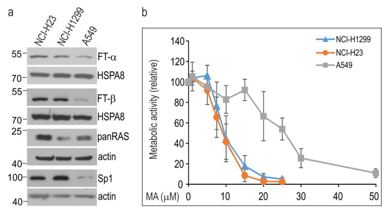
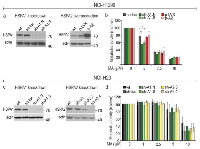
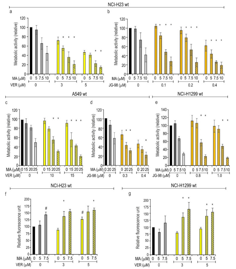
Manumycin A (MA) is a well-tolerated natural antibiotic showing pleiotropic anticancer effects in various preclinical in vitro and in vivo models. Anticancer drugs may themselves act as stressors to induce the cellular adaptive mechanism that can minimize their cytotoxicity. Heat shock proteins (HSPs) as cytoprotective factors can counteract the deleterious effects of various stressful stimuli. In this study, we examined whether the anticancer effects of MA can be counteracted by the mechanism related to HSPs belonging to the HSPA (HSP70) family. We found that MA caused cell type-specific alterations in the levels of HSPAs. These changes included concomitant upregulation of the stress-inducible (HSPA1 and HSPA6) and downregulation of the non-stress-inducible (HSPA2) paralogs. However, neither HSPA1 nor HSPA2 were necessary to provide protection against MA in lung cancer cells. Conversely, the simultaneous repression of several HSPA paralogs using pan-HSPA inhibitors (VER-155008 or JG-98) sensitized cancer cells to MA. We also observed that genetic ablation of the heat shock factor 1 (HSF1) transcription factor, a main transactivator of HSPAs expression, sensitized MCF7 cells to MA treatment. Our study reveals that inhibition of HSF1-mediated heat shock response (HSR) can improve the anticancer effect of MA. These observations suggest that targeting the HSR- or HSPA-mediated adaptive mechanisms may be a promising strategy for further preclinical developments.

Keywords: HSP70; HSPA inhibitors; HSPA1; HSPA2; breast cancer; heat shock factor 1; heat shock proteins; lung cancer; manumycin A.


Conflict of interest statement

The authors declare no conflict of interest.

Figures

Figure 1
Figure 1
NSCLC cell lines show different sensitivity to manumycin A (MA). (a) The basal levels of MA targets in NSCLC cells. Representative immunoblots are shown (n ≥ 3); actin or HSPA8 were used as a protein loading control. Numbers on the left or right side of the blots indicate molecular weight of a protein size marker. (b) Dose-response curves of MA in NSCLC cell lines. Cell viability was measured following 72 h treatment with MA (0–50 µM) using an MTS assay. Results (mean ± SD from at least four independent measurements, each in three technical replicates) are expressed relatively to untreated control.
Figure 2
Figure 2
MA-induced changes in (a) the protein level of the HSPA paralogs, and (b) HSF1 phosphorylation in lung cancer cells. Levels of HSPA paralogs (a); total HSF1 (b); phosphorylated HSF1 (b) in cells non-treated or treated with MA for 24 hours (in (a)) or for 1–9 hours (h) (in (b)). Representative immunoblots are shown (n ≥ 3); actin was used as a protein loading control. Numbers on the left or right side of the immunoblot indicate the molecular weight of a protein size marker.
Figure 3
Figure 3
Manipulations in the protein levels of HSPA1 and HSPA2 have limited effect on sensitivity of NSCLC cells to manumycin A (MA). (a,c) Immunoblots showing levels of HSPA1 and HSPA2 in wild-type (wt) and lentivirally-modified cells; sh-luc control cells were transduced with a non-targeting shRNA-luc sequence; sh-A1.N and sh-A1.S cell lines were transduced with HSPA1-targeting shRNA sequences; control p-LVX cells were transduced with lentiviruses bearing the “empty” pLVX-Puro vector; p-A2 cells were transduced with pLVX-Puro plasmid encoding HSPA2 protein under the control of the CMV promoter; sh-A2.3 and sh-A2.4 cell lines were transduced with HSPA2-targeting shRNA sequences. Representative immunoblots are shown (n ≥ 3); actin was used as a protein loading control. These model cell lines were described in detail previously [26,27]. (b,d) Cell viability was measured using MTS assay after 72 h treatment with MA (0–10 µM). Results are expressed as mean ± SD in relation to untreated control (n = 4, each in three technical replicates, * p < 0.05, statistical significance was determined by two-tailed t-test). The numbers on the right side of immunoblots indicate molecular weight of the protein size marker.
Figure 4
Figure 4
Effects of single or combined treatment (72 h) with (a,c,f,g) VER-155008 (VER) and manumycin A (MA) or (b,d,e) JG-98 and MA. Cell viability (ae) was measured using MTS assay. Results are expressed in relation to the untreated control (mean ± SD, n ≥ 3, each in triplicate). Cytotoxicity (f,g) was evaluated using the CellTox™ cytotoxicity assay (mean ± SD, n ≥ 3, each in duplicate). Statistical significance was determined using two-tailed t-test; * p < 0.05, differences were considered significant if the value obtained for a double treatment was significantly different (p < 0.05) from the value obtained for each of the single treatments; # p < 0.05, statistical significance determined for single treatments (f,g).
Figure 5
Figure 5
Effects of HSF1 knockout on response of MCF7 cells to manumycin A (MA). (a) Levels of total HSF1 and its phosphorylation at the serine 326 residue in cells exposed to MA for 0–9 h. (b) Levels of HSPA proteins in control and HSF1-null cells. (c) Viability of wild type (wt) cells after single and double treatment (72 h) with MA (0–5 µM) and/or VER-155008 (VER) (10 µM) assessed using an MTS assay. Results (mean ± SD; n = 5, each in two technical replicates) are expressed in relation to values obtained for non-treated cells. (d) Levels of HSF1 in wild-type (wt); control (CRISPR-CTL, a pool of non-edited isogenic clones); and two HSF1-null isogenic clones (CRISPR-K14, CRISPR-K45). (e) Levels of HSPA proteins in non-treated and MA-treated control and HSF1-null cells. (f) Viability of control and HSF1-null cells after MA (0–5 µM) treatment (72 h) assessed using the alamarBlue® assay. Results (mean ± SD; n = 4, each in two technical replicates) are expressed in relation to values obtained for non-treated cells. (g) Viability of control and HSF1-null cells after single and double treatment (72 h) with MA (0–5 µM) and/or VER-155008 (VER) (10 µM) assessed using an MTS assay. Results (mean ± SD; n = 5, each in two technical replicates) are expressed in relation to values obtained for non-treated cells, control, and HSF1-null cells. In (a,b,d,e) representative immunoblots are shown (n = 3); actin was used as a protein loading control. Statistical significance was determined by two-tailed t-test. The numbers on the left side of blots indicate molecular weight of the protein size marker. In (c,g) * p < 0.05, differences were considered statistically significant if the value obtained for a double treatment was significantly different (p < 0.05) from the value obtained for each of the single treatments. In (f) * p < 0.05, statistically significant differences in relation to control CRISPR-CTL cells. In (g) # p < 0.05, statistically significant differences in relation to CRISPR-CTL cells, and & = 0.053.

References

    1. Bérdy J. Bioactive Microbial Metabolites. J. Antibiot. 2005;58:1–26. doi: 10.1038/ja.2005.1. - DOI - PubMed
    1. Takahashi Y., Nakashima T. Actinomycetes, an Inexhaustible Source of Naturally Occurring Antibiotics. Antibiotics. 2018;7:45. doi: 10.3390/antibiotics7020045. - DOI - PMC - PubMed
    1. Hara M., Akasaka K., Akinaga S., Okabe M., Nakano H., Gomez R., Wood D., Uh M., Tamanoi F. Identification of Ras farnesyltransferase inhibitors by microbial screening. Proc. Natl. Acad. Sci. USA. 1993;90:2281–2285. doi: 10.1073/pnas.90.6.2281. - DOI - PMC - PubMed
    1. Ito T., Kawata S., Tamura S., Igura T., Nagase T., Miyagawa J.I., Yamazaki E., Ishiguro H., Matasuzawa Y. Suppression of human pancreatic cancer growth in BALB/c nude mice by manumycin, a farnesyl:protein transferase inhibitor. Jpn. J. Cancer Res. 1996;87:113–116. doi: 10.1111/j.1349-7006.1996.tb03146.x. - DOI - PMC - PubMed
    1. She M., Yang H., Sun L., Yeung S.C. Redox control of manumycin A-induced apoptosis in anaplastic thyroid cancer cells: Involvement of the xenobiotic apoptotic pathway. Cancer Biol. Ther. 2006;5:275–280. doi: 10.4161/cbt.5.3.2383. - DOI - PubMed

Publication types

MeSH terms