. 2021 Jun 7;7(6):454.
doi: 10.3390/jof7060454.

Characterisation of Aspergillus fumigatus Endocytic Trafficking within Airway Epithelial Cells Using High-Resolution Automated Quantitative Confocal Microscopy

Affiliations

Characterisation of Aspergillus fumigatus Endocytic Trafficking within Airway Epithelial Cells Using High-Resolution Automated Quantitative Confocal Microscopy

Nagwa Ben-Ghazzi et al. J Fungi (Basel). .

Abstract

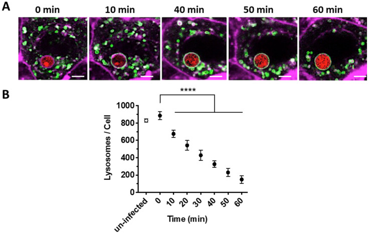
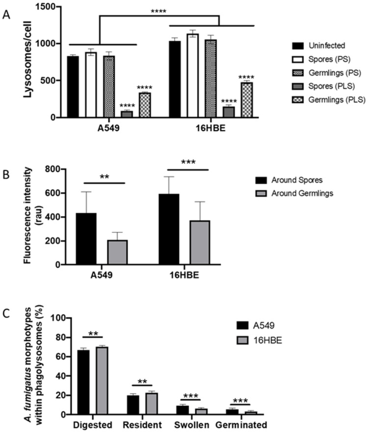
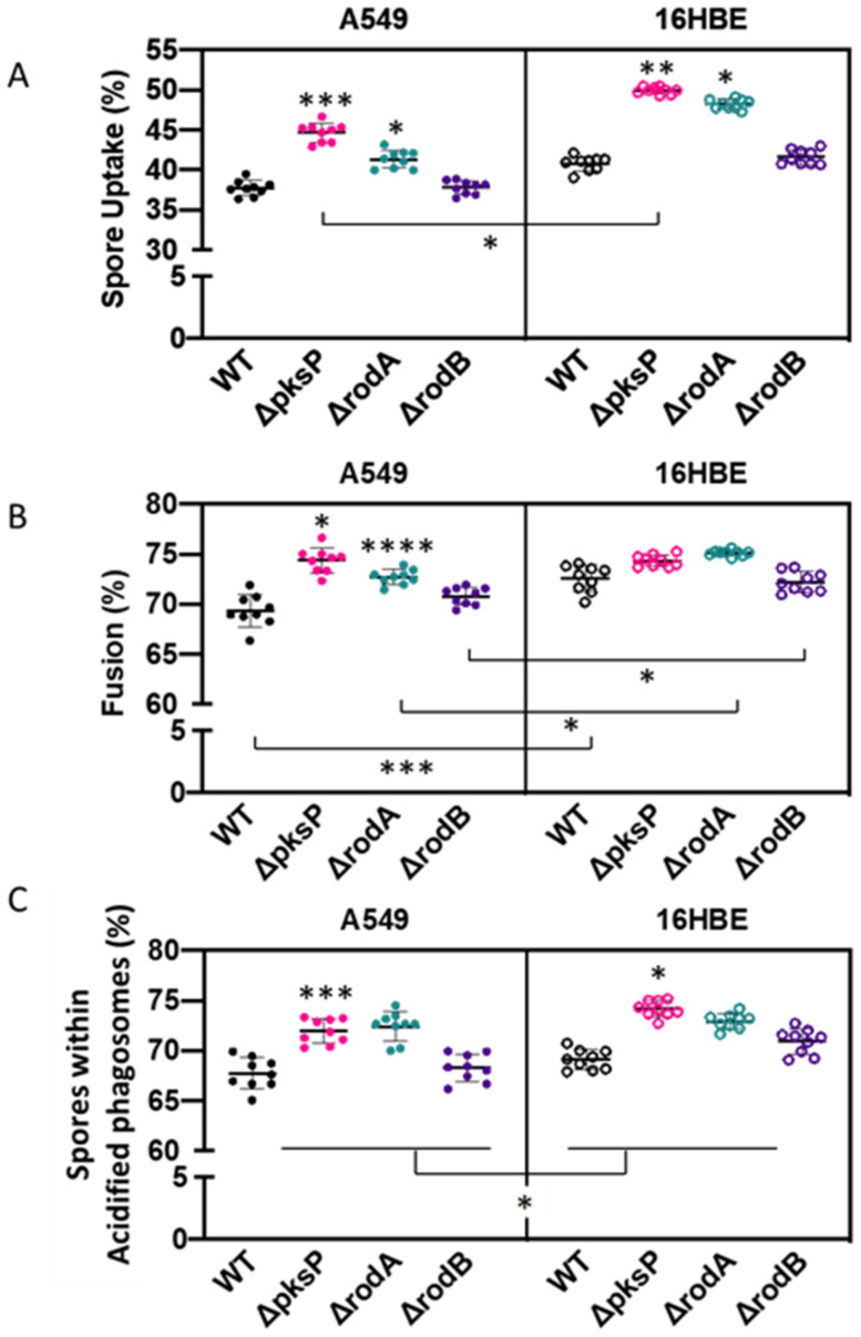
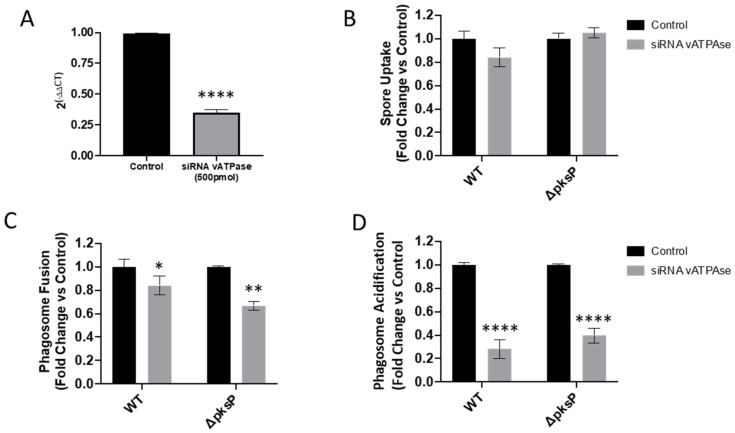
The precise characterization of the mechanisms modulating Aspergillus fumigatus survival within airway epithelial cells has been impaired by the lack of live-cell imaging technologies and user-friendly quantification approaches. Here we described the use of an automated image analysis pipeline to estimate the proportion of A. fumigatus spores taken up by airway epithelial cells, those contained within phagolysosomes or acidified phagosomes, along with the fungal factors contributing to these processes. Coupling the use of fluorescent A. fumigatus strains and fluorescent epithelial probes targeting lysosomes, acidified compartments and cell membrane, we found that both the efficacy of lysosome recruitment to phagosomes and phagosome acidification determines the capacity of airway epithelial cells to contain A. fumigatus growth. Overall, the capability of the airway epithelium to prevent A. fumigatus survival was higher in bronchial epithelial than alveolar epithelial cells. Certain A. fumigatus cell wall mutants influenced phagosome maturation in airway epithelial cells. Taken together, this live-cell 4D imaging approach allows observation and measurement of the very early processes of A. fumigatus interaction within live airway epithelial monolayers.

Keywords: Aspergillus fumigatus; airway epithelial cells; phagocytosis.


Conflict of interest statement

In the past five years, S.G. has received research funds from Pfizer and has been a council member of the International Society of Human and Animal Mycology (ISHAM). Denning and family hold Founder shares in F2G Ltd, a University of Manchester spin-out antifungal discovery company. He acts, or has recently acted, as a consultant to Pulmatrix, Pulmocide, Zambon, iCo Therapeutics, Mayne Pharma, Biosergen, Bright Angel Therapeutics, Cipla and Metis. He sits on the DSMB for a SARS CoV2 vaccine trial. In the last three years, he has been paid for talks on behalf of Dynamiker, Hikma, Gilead, Merck, Mylan and Pfizer. In the last 5 years D.T has received research funds from Gilead Science and has acted as a consultant for OwlStone Medical.

Figures

Figure 1
Figure 1
Dynamics of phagosome-lysosome fusion observed during A. fumigatus phagocytosis. (A) Representative time-lapse confocal images from the central focal plane of a single A. fumigatus spore (red) engulfed and localized within A549 alveolar epithelial cell monolayer (magenta) from 5 to 6 h post-infection in 10 min intervals. (B) Number of lysosomes within airway epithelial cells which have taken up A. fumigatus spores is reduced over time. (mean ± SD of three biological replicates assessed in technical triplicates). Scale Bar = 5 μm. (**** p < 0.0001, compared to 0 min). Magenta = epithelial cells membrane (CellMask Deep Red), Green = lysosomes (GFP-LAMP), red = A. fumigatus (MFIGRag29, Table S1).
Figure 2
Figure 2
A. fumigatus fate within phagolysosomes of A549 alveolar epithelial cells at 18 h post-infection. (A) Digested spore (asterisk). (B) Resting (white arrow) and swollen spore (yellow arrow) inside the phagolysosome. (C) Germinated spore within phagolysosome. Scale Bar = 5 μm. Magenta = airway epithelial cells membrane (CellMask Deep Red), Green = lysosomes (GFP-LAMP), red = A. fumigatus (MFIGRag29, Table S1).
Figure 3
Figure 3
Suboptimal lysosome fusion permits A. fumigatus germination within the airway epithelium. (A) Proportion of A. fumigatus spores and germlings within A549 alveolar epithelial cells. (B) Proportion of internalized spores and germlings within phagolysosomes of A549 cells. (C) Proportion of internalized spores and germlings within unfused phagosomes of A549 cells. Data represents mean and standard deviation of five biological replicates. Differences in proportions between time points for spores and germlings were independently tested using the Fischer Exact Test. **** p < 0.0001.
Figure 4
Figure 4
Killing of A. fumigatus within A549 alveolar epithelial cell lines. (A) Single airway epithelial cell containing digested (triangles) and killed germlings (asterisks) within phagolysosomes. The cross symbol refers to the crescent feature after digestion of a spore within a phagosome. (B) Hyphal killing occurs within phagolysosomes (yellow arrows) and within phagosomes (indicated by dashed circle). (C) Killing of A. fumigatus within the acidified phagosome. (D) Residence of A. fumigatus spore co-localized within acidic niche for more than 18 h. (E) Spore swelling within the acidified phagosome 24 h of inoculation. Color code for panels A and B: Magenta = epithelial cells membrane (CellMask Deep Red), Green = lysosomes (GFP-LAMP1), red = A. fumigatus (MFIGRag29). Color code for panels C–E: Magenta = epithelial cells membrane (CellMask Deep Red), Red = Acidic organelles (RFP-Lysotracker), Green = A. fumigatus (MFIGGFP4, Table S1). Scale Bar = 5 μm.
Figure 5
Figure 5
Phagolysosome formation is critical to prevent A. fumigatus growth within airway epithelial cells. (A) Changes in the number of lysosomes per cell in 16HBE and A549 lung epithelial cells challenged and unchallenged with A. fumigatus spores. Asterisks represent differences between spores or germlings compared with uninfected for each cell line. Lines represent differences between groups for each cell line. (B) Intensity of relative LAMP1-GFP fluorescence intensity around spore or germlings-containing phagolysosomes of A549 and 16HBE lung epithelial cells. Asterisks represent differences between spores and gemlings for each cell line. (C) Fate of A. fumigatus spore within phagosomes (PS) and phagolysosomes (PLS) of A549 and 16HBE lung epithelial cells. Asterisks represent differences for each morphotype between cell lines. Data represents mean and standard deviation of three biological replicates assayed in technical triplicates. (** p < 0.01; *** p < 0.001; **** p < 0.0001).
Figure 6
Figure 6
Impact of A. fumigatus cell wall mutants in (A) spore uptake, (B) phagolysosome fusion and (C) phagosome acidification of A549 and 16HBE airway epithelial cells at 3 h post-infection. Asterisks represent differences vs. wild type for each mutant within cell lines. Lines represent differences between cell lines for each mutant. Data shown as mean ± standard deviation of three biological replicates assayed in technical triplicates (* p < 0.05, ** p < 0.01, *** p < 0.001, **** p < 0.0001).
Figure 7
Figure 7
Impact of vATPase siRNA silencing of A549 alveolar epithelial cells in (A) vATPAse gene expression, (B) A. fumigatus spore uptake, (C) phagolysosome fusion and (D) phagosome acidification by A549 and 16HBE cells at 3 h post infection. Asterisks represent differences vs. control. Data shown as mean ± standard deviation of three biological and technical triplicates (* p < 0.05, ** p <0.01, **** p <0.0001).

Similar articles

Cited by

References

    1. Bongomin F., Gago S., Oladele R.O., Denning D.W. Global and multi-national prevalence of fungal diseases—Estimate precision. J. Fungi. 2017;3:57. doi: 10.3390/jof3040057. - DOI - PMC - PubMed
    1. Brown G.D., Denning D.W., Gow N.A.R., Levitz S.M., Netea M.G., White T.C. Hidden killers: Human fungal infections. Sci. Transl. Med. 2012;4:165rv13. doi: 10.1126/scitranslmed.3004404. - DOI - PubMed
    1. Latge J.P., Chamilos G. Aspergillus fumigatus and Aspergillosis in 2019. Clin. Microbiol. Rev. 2019;33:e00140-18. doi: 10.1128/CMR.00140-18. - DOI - PMC - PubMed
    1. Van De Veerdonk F.L., Gresnigt M.S., Romani L., Netea M.G., Latgé J.P. Aspergillus fumigatus morphology and dynamic host interactions. Nat. Rev. Microbiol. 2017;15:661–674. doi: 10.1038/nrmicro.2017.90. - DOI - PubMed
    1. Heinekamp T., Schmidt H., Lapp K., Pähtz V., Shopova I., Köster-Eiserfunke N., Krüger T., Kniemeyer O., Brakhage A.A. Interference of Aspergillus fumigatus with the immune response. Semin. Immunopathol. 2015;37:141–152. doi: 10.1007/s00281-014-0465-1. - DOI - PMC - PubMed

LinkOut - more resources