Review
. 2021 Jun 7;22(11):6158.
doi: 10.3390/ijms22116158.

Roles of Key Ion Channels and Transport Proteins in Age-Related Hearing Loss

Affiliations
Review

Roles of Key Ion Channels and Transport Proteins in Age-Related Hearing Loss

Parveen Bazard et al. Int J Mol Sci. .

Abstract

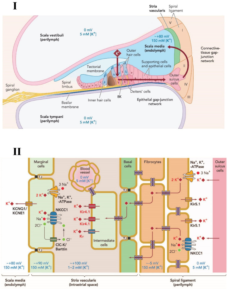
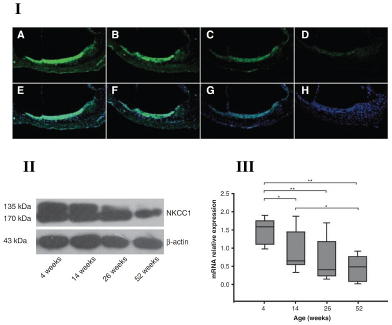
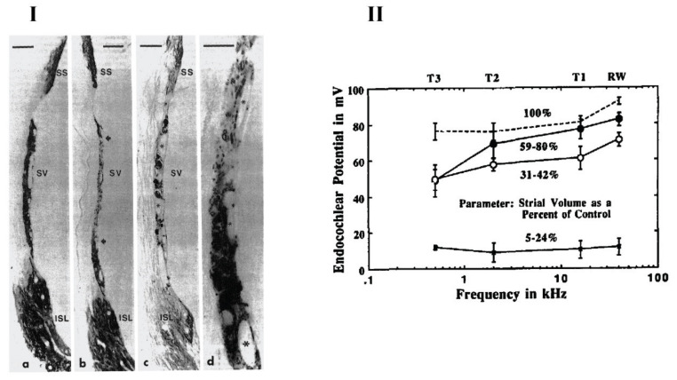
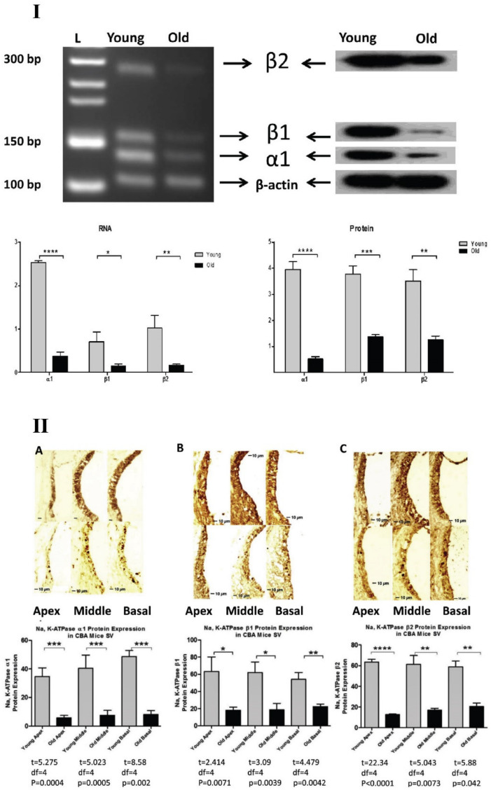
The auditory system is a fascinating sensory organ that overall, converts sound signals to electrical signals of the nervous system. Initially, sound energy is converted to mechanical energy via amplification processes in the middle ear, followed by transduction of mechanical movements of the oval window into electrochemical signals in the cochlear hair cells, and finally, neural signals travel to the central auditory system, via the auditory division of the 8th cranial nerve. The majority of people above 60 years have some form of age-related hearing loss, also known as presbycusis. However, the biological mechanisms of presbycusis are complex and not yet fully delineated. In the present article, we highlight ion channels and transport proteins, which are integral for the proper functioning of the auditory system, facilitating the diffusion of various ions across auditory structures for signal transduction and processing. Like most other physiological systems, hearing abilities decline with age, hence, it is imperative to fully understand inner ear aging changes, so ion channel functions should be further investigated in the aging cochlea. In this review article, we discuss key various ion channels in the auditory system and how their functions change with age. Understanding the roles of ion channels in auditory processing could enhance the development of potential biotherapies for age-related hearing loss.

Keywords: age-related hearing loss; aging; auditory; cochlea; deafness; hearing; inner ear; ion channels; potassium channels; presbycusis.


Conflict of interest statement

The authors declare no conflict of interest.

Figures

Figure 1
Figure 1
Schematic diagram of potassium circulation, ion channels and transporters in the cochlear lateral wall. (I) Potassium exits the hair cell and re-circulates into the endolymph of the scala media via various structures and ion channels in the supporting cells and lateral wall of the cochlea. (II) Various ion transporter channels; NKCC1; Na, K-ATPase; KCNQ1; Kir 4.1; Kir 5.1 and Kv, are expressed in the stria vascularis and spiral ligament that participate in potassium circulation and endocochlear potential generation. TJ: Tight Junctions. NKCC1, a key transporter in the cochlea, is expressed in spiral ligament and stria vascularis cells. Adapted from Hibino and Kurachi (2006) [25], with permission from the publisher.
Figure 2
Figure 2
Aging decline of NKCC1 (sodium-potassium-chloride cotransporter) in C57BL/6J mouse cochlear lateral wall with four different age-group animals; 4 weeks old, 14 weeks old, 26 weeks old and 52 weeks old. (I) Age-related changes of NKCC1 protein stain with FITC (Fluorescein isothiocyanate, green fluorescence—top row) and the nuclei counter stained by DAPI (4′,6-diamidino-2-phenylindole, blue fluorescence—bottom row) for 4 animal groups at 400× magnification; A, E—4-week-old group; B,F—14-week-old group; C, G—26-week-old group and D, H—52-week-old group. (II,III) Western blotting and real-time polymerase chain reaction (RT-PCR) results from the four different age groups showed the gradual reduction in NKCC1 at both protein and gene expression levels. * p < 0.05, ** p < 0.01. From Liu et al. (2014) [55], with permission from the publisher.
Figure 3
Figure 3
Na, K-ATPase expression in the cochlear lateral wall and endocochlear potential recordings for different age-group gerbils. (I) Middle turn sections of gerbil cochlea showing the Na, K-ATPase staining for a 6-month-old gerbil (a) and a 36-month-old gerbil (b–d). Various parts of lateral wall were stained for Na, K-ATPase expression; marginal cells in stria vascularis (SV), fibrocytes in the suprastrial zone (SS) and the inferior spiral ligament (ISL). There was a reduction in NA, K-ATPase immunostaining in the central portion of the SV (b—between arrows) as well as fibrocytes in the SS zone and ISL (c). Higher magnification of strial atrophy in part (b) is depicted in part (d). Strial capillaries (asterisks) were present in old age gerbil cochlea and remain patent (c and d). Scale Bars: a, b, c = 20 μM and d = 10 μM. Magnifications: a, b and c = ×800 and d = ×1800. (II) Mean values of endocochlear potentials of nine young animals (dotted line) and three groups of 35- to 38-months-old animals. The measurements were done at four positions—round window and three turns; T1, T2 and T3. Old animals were grouped based on the percentage of Na, K-ATPase immunostaining remaining as compared to the young adult control group. From Schulte and Schmiedt (1992) [118], with permission from the publisher.
Figure 4
Figure 4
Age-related decrease in Na, K-ATPase subunits in CBA/CaJ mouse cochlea. (I) Protein lysate and mRNA extracts were analyzed for young adult (3 months) and old (30 months) CBA/CaJ mice using western blotting and RT-PCR techniques, respectively. There was significant decrease in α1, β1, β2 subunits of Na, K-ATPase at both protein and gene expression levels. (II) Cross sections of cochlea with immunostaining further confirmed the results using immunohistochemistry i.e., there was a significant decrease for Na, K-ATPase subunits α1, β1, β2 with aging in all three cochlear turns. * p < 0.05, ** p < 0.01, *** p < 0.005, **** p < 0.001. From Ding et al. (2018) [121], with permission from the publisher.
Figure 5
Figure 5
Age-related decrease of Kir 5.1 channels in the cochlear lateral wall of C57BL/6J mice. Cochlea from 4 different age-group C57BL/6J mice were analyzed using three different techniques; immunohistochemistry (I) scale bar = 10 µm, western blotting, (II), and RT-PCR (III). Four age groups tested: 4-week-old group, 14-week-old group, 24-week-old group, and 52-week-old group. There was a steady decrease in Kir 5.1 expressions levels at both protein and gene levels with age. * p < 0.05, ** p < 0.01. From Pan et al. (2016) [193], with permission from the publisher.

References

    1. Frisina D.R., Frisina R.D. Speech recognition in noise and presbycusis: Relations to possible neural mechanisms. Hear. Res. 1997;106:95–104. doi: 10.1016/S0378-5955(97)00006-3. - DOI - PubMed
    1. Dalton D.S., Cruickshanks K.J., Klein B.E., Klein R., Wiley T.L. The Impact of Hearing Loss on Quality of Life in Older Adults. Gerontologist. 2003;43:661–668. doi: 10.1093/geront/43.5.661. - DOI - PubMed
    1. Huang Q., Tang J. Age-related hearing loss or presbycusis. Eur. Arch. Oto-Rhino-Laryngol. 2010;267:1179–1191. doi: 10.1007/s00405-010-1270-7. - DOI - PubMed
    1. Kramer S.E., Kapteyn T.S., Kuik D.J. The Association of Hearing Impairment and Chronic Diseases with Psychosocial Health Status in Older Age. J. Aging Health. 2002;14:122–137. doi: 10.1177/089826430201400107. - DOI - PubMed
    1. Lin F.R., Ferrucci L., Metter E.J., An Y., Zonderman A.B. Hearing loss and cognition in the Baltimore Longitudinal Study of Aging. Neuropsychology. 2011;25:763–770. doi: 10.1037/a0024238. - DOI - PMC - PubMed