

Ensemble Method of Convolutional Neural Networks with Directed Acyclic Graph Using Dermoscopic Images: Melanoma Detection Application
- PMID: 34200521
- PMCID: PMC8229112
- DOI: 10.3390/s21123999
Ensemble Method of Convolutional Neural Networks with Directed Acyclic Graph Using Dermoscopic Images: Melanoma Detection Application
Abstract
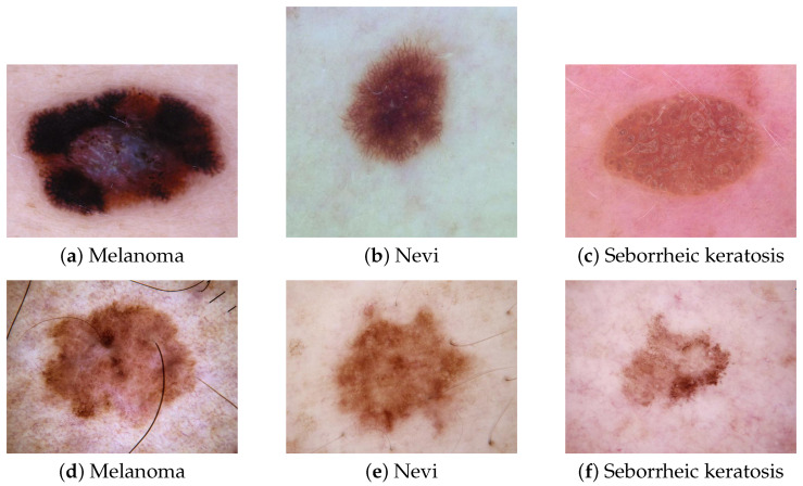
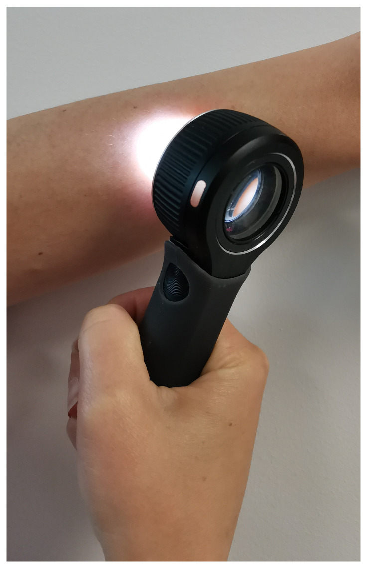
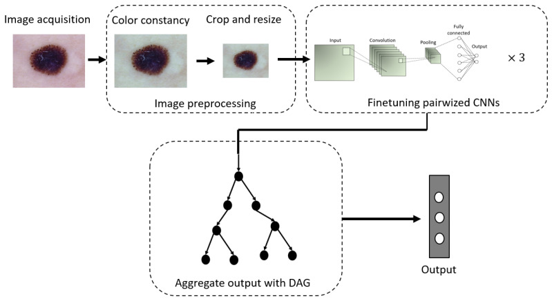
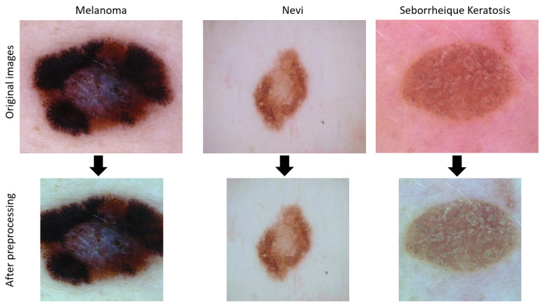
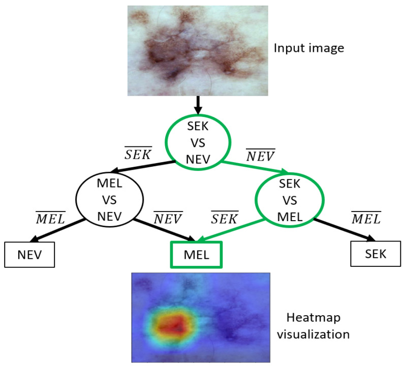
The early detection of melanoma is the most efficient way to reduce its mortality rate. Dermatologists achieve this task with the help of dermoscopy, a non-invasive tool allowing the visualization of patterns of skin lesions. Computer-aided diagnosis (CAD) systems developed on dermoscopic images are needed to assist dermatologists. These systems rely mainly on multiclass classification approaches. However, the multiclass classification of skin lesions by an automated system remains a challenging task. Decomposing a multiclass problem into a binary problem can reduce the complexity of the initial problem and increase the overall performance. This paper proposes a CAD system to classify dermoscopic images into three diagnosis classes: melanoma, nevi, and seborrheic keratosis. We introduce a novel ensemble scheme of convolutional neural networks (CNNs), inspired by decomposition and ensemble methods, to improve the performance of the CAD system. Unlike conventional ensemble methods, we use a directed acyclic graph to aggregate binary CNNs for the melanoma detection task. On the ISIC 2018 public dataset, our method achieves the best balanced accuracy (76.6%) among multiclass CNNs, an ensemble of multiclass CNNs with classical aggregation methods, and other related works. Our results reveal that the directed acyclic graph is a meaningful approach to develop a reliable and robust automated diagnosis system for the multiclass classification of dermoscopic images.
Keywords: computer-aided system; deep learning; dermoscopic images; directed acyclic graph; ensemble method; fusion-based model; melanoma detection; multiclass classification; skin cancer.
Conflict of interest statement
The authors declare no conflict of interest.
Figures
Similar articles
-
Computer Aided Diagnosis of Melanoma Using Deep Neural Networks and Game Theory: Application on Dermoscopic Images of Skin Lesions.Int J Mol Sci. 2022 Nov 10;23(22):13838. doi: 10.3390/ijms232213838. Int J Mol Sci. 2022. PMID: 36430315 Free PMC article.
-
Skin lesion classification with ensembles of deep convolutional neural networks.J Biomed Inform. 2018 Oct;86:25-32. doi: 10.1016/j.jbi.2018.08.006. Epub 2018 Aug 10. J Biomed Inform. 2018. PMID: 30103029
-
Fusing fine-tuned deep features for skin lesion classification.Comput Med Imaging Graph. 2019 Jan;71:19-29. doi: 10.1016/j.compmedimag.2018.10.007. Epub 2018 Nov 3. Comput Med Imaging Graph. 2019. PMID: 30458354
-
Deep Learning Approaches Towards Skin Lesion Segmentation and Classification from Dermoscopic Images - A Review.Curr Med Imaging. 2020;16(5):513-533. doi: 10.2174/1573405615666190129120449. Curr Med Imaging. 2020. PMID: 32484086 Review.
-
Skin cancer classification via convolutional neural networks: systematic review of studies involving human experts.Eur J Cancer. 2021 Oct;156:202-216. doi: 10.1016/j.ejca.2021.06.049. Epub 2021 Sep 8. Eur J Cancer. 2021. PMID: 34509059
Cited by
-
Analysis of Artificial Intelligence-Based Approaches Applied to Non-Invasive Imaging for Early Detection of Melanoma: A Systematic Review.Cancers (Basel). 2023 Sep 23;15(19):4694. doi: 10.3390/cancers15194694. Cancers (Basel). 2023. PMID: 37835388 Free PMC article. Review.
-
The role of artificial intelligence and convolutional neural networks in the management of melanoma: a clinical, pathological, and radiological perspective.Melanoma Res. 2024 Apr 1;34(2):96-104. doi: 10.1097/CMR.0000000000000951. Epub 2023 Dec 22. Melanoma Res. 2024. PMID: 38141179 Free PMC article.
-
Computer Aided Diagnosis of Melanoma Using Deep Neural Networks and Game Theory: Application on Dermoscopic Images of Skin Lesions.Int J Mol Sci. 2022 Nov 10;23(22):13838. doi: 10.3390/ijms232213838. Int J Mol Sci. 2022. PMID: 36430315 Free PMC article.
-
Multi-Class Skin Lesions Classification Using Deep Features.Sensors (Basel). 2022 Oct 29;22(21):8311. doi: 10.3390/s22218311. Sensors (Basel). 2022. PMID: 36366009 Free PMC article.
-
Detection of Skin Cancer Based on Skin Lesion Images Using Deep Learning.Healthcare (Basel). 2022 Jun 24;10(7):1183. doi: 10.3390/healthcare10071183. Healthcare (Basel). 2022. PMID: 35885710 Free PMC article.
References
-
- Leiter U., Keim U., Garbe C. Sunlight, Vitamin D and Skin Cancer. Springer; Cham, Switzerland: 2020. Epidemiology of skin cancer: Update 2019; pp. 123–139. - PubMed
-
- Grob J.J., Gaudy C. Early detection of melanoma. Discussing a new approach. Rev. Prat. 2007;57:1397–1400. - PubMed
-
- Codella N., Rotemberg V., Tschandl P., Celebi M.E., Dusza S., Gutman D., Helba B., Kalloo A., Liopyris K., Marchetti M., et al. Skin lesion analysis toward melanoma detection 2018: A challenge hosted by the international skin imaging collaboration (isic) arXiv. 20191902.03368
MeSH terms
LinkOut - more resources
Full Text Sources
Other Literature Sources
Medical
Miscellaneous
