Review
. 2021 Jun 10;10(6):1454.
doi: 10.3390/cells10061454.

Murder on the Ovarian Express: A Tale of Non-Autonomous Cell Death in the Drosophila Ovary

Affiliations
Review

Murder on the Ovarian Express: A Tale of Non-Autonomous Cell Death in the Drosophila Ovary

Diane Patricia Vig Lebo et al. Cells. .

Abstract

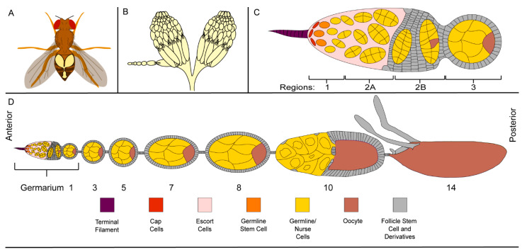
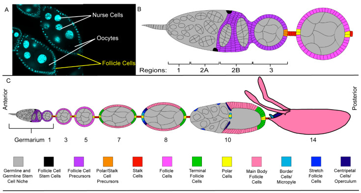
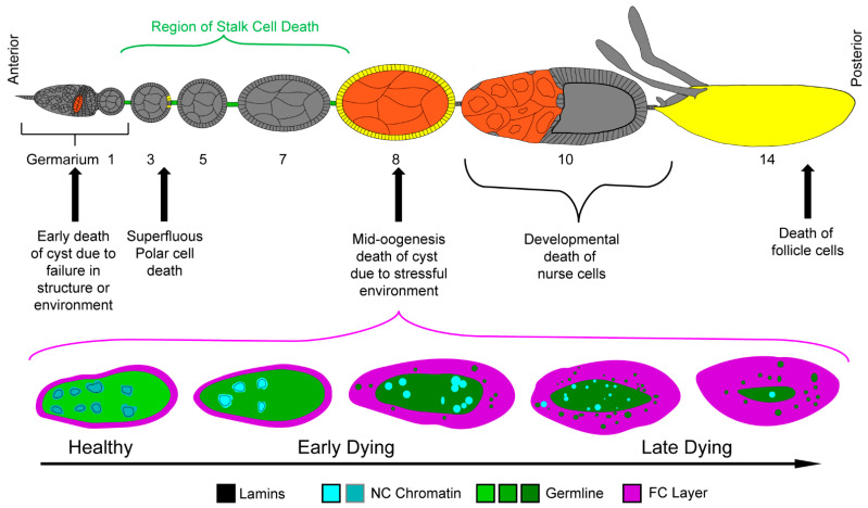
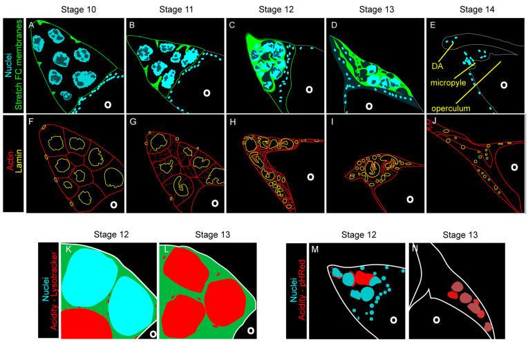
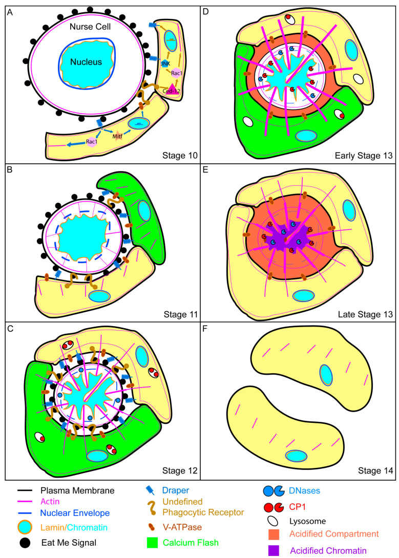
Throughout oogenesis, Drosophila egg chambers traverse the fine line between survival and death. After surviving the ten early and middle stages of oogenesis, egg chambers drastically change their size and structure to produce fully developed oocytes. The development of an oocyte comes at a cost, the price is the lives of the oocyte's 15 siblings, the nurse cells. These nurse cells do not die of their own accord. Their death is dependent upon their neighbors-the stretch follicle cells. Stretch follicle cells are nonprofessional phagocytes that spend the final stages of oogenesis surrounding the nurse cells and subsequently forcing the nurse cells to give up everything for the sake of the oocyte. In this review, we provide an overview of cell death in the ovary, with a focus on recent findings concerning this phagocyte-dependent non-autonomous cell death.

Keywords: Drosophila; cell corpse clearance; efferocytosis; oogenesis; ovary; phagocytosis; phagoptosis.


Conflict of interest statement

The authors declare no conflict of interest.

Figures

Figure 1
Figure 1
Structure of the Drosophila ovary (A) The relative size and position of ovaries within a Drosophila female. (B) A schematic of Drosophila ovaries showing an ovariole and its egg chambers. (C) The structure of a germarium with each of the germline cells, stem cells and niche in color and somatic cells in grey (see color scheme in Figure 1D). The terminal filament, cap cells, and escort cells found in region 1 act as the germline stem cell niche. Each egg chamber in the ovariole is derived, in part, from one of the germline stem cells. (D) A detailed schematic of an ovariole with the germline cells, stem cells, and niche in color and somatic cells in grey. An ovariole is composed of several egg chambers at different stages of development.
Figure 2
Figure 2
Somatic Cells of the Drosophila ovary (A) DAPI-stained egg chambers containing 15 NCs and a single oocyte. This syncytium of 16 cells is surrounded by a layer of smaller epithelial FCs. (B) A detailed schematic of an ovariole with the somatic cells in color and the germline cells, stem cells, and niche in grey (see color scheme in Figure 2C). The FCs are produced by the follicle cell stem cells found in region 2A of the germarium. (C) A detailed schematic of an ovariole with the somatic cells in color and the germline cells, stem cells, and niche in grey.
Figure 3
Figure 3
Cell death in oogenesis. Top—Schematic of ovariole with germline cell death events highlighted in orange and somatic cell death events highlighted in yellow and green. Bottom—Phases of cell death in egg chambers during mid-oogenesis are illustrated. Healthy egg chambers contain NCs with dispersed chromatin and intact nuclear lamina. The FC layer (magenta) surrounding the syncytium is thin, just 1 cell thick. As the germline dies, the chromatin condenses (as shown by the increasing brightness of the cyan) and the lamins are cleaved and become cytoplasmic (as shown by the germline green becoming darker). The chromatin condenses and fragments as germline cell death progresses. The follicle cells then efferocytose the germline material (green vesicles) to clear it away.
Figure 4
Figure 4
Developmental death of the nurse cells is orchestrated by the stretch follicle cells. (AE) Images of the anterior portion of egg chambers during the late stages of oogenesis. (Adapted from [29]). (A) The anterior end of a stage 10 egg chamber with dispersed NC nuclei and surrounding FCs. (B) In a stage 11 egg chamber, NCs become smaller as they dump their cytoplasmic contents into the oocyte. Soon after, the stretch FCs invade the space between the NCs. (C) By stage 12, all of the cytoplasmic contents have been dumped into the oocyte and the stretch FCs have completely surrounded the NCs. (D) During stage 13, the NCs start to disappear and the dorsal appendages (DA) start to grow. (E) By stage 14, the NCs are completely eliminated, the DAs have fully extended, and the micropyle and operculum are complete. (FJ) Actin and lamin dynamics during developmental nurse cell death. (Adapted from [75]). (F) During stage 10, the only visible F-actin is cortical. The NC lamina, however, have already begun to fold creating crenellations (as compared to the smooth lamina of the stretch FCs). (GI) During stages 11–13, actin bundles extend into the nucleus and the lamina undergoes additional folding, forming involutions and focal disruptions. (J) By stage 14, the only lamins present are those of the stretch FCs. The actin again can only be found subcortically. (K,L) NC nuclei become acidified during stages 12 and 13 (Adapted from [31]). (K) During stage 12, acidic organelles in the stretch FCs can be seen surrounding the NCs. (L) During stage 13, NCs become acidified. (M,N) NC acidity originates in the stretch FCs (Adapted from [31]). (M) During stage 12, acidity can be seen at the periphery of the nurse cells, near the stretch FCs. (N) During stage 13, acidity has flooded into the rest of the NC remnant. (KN) The oocyte (O) is not affected by acidity.
Figure 5
Figure 5
A model of developmental death and clearance of the nurse cells. (A) NCs of stage 10 egg chambers are at the cusp of death. The NC nucleus is already starting to fold, but the only visible actin is cortical. The NC has exposed a hypothetical eat me signal which is recognized by the phagocytic receptors on the FC membranes. When the Ced-12 pathway is triggered, it likely signals through Rac1 to the JNK pathway to the nucleus to increase production of a variety of proteins including Drpr. Activation of the Drpr pathway may send signals to the nucleus via MITF to produce V-ATPases. Drpr and Ced-12 also signal Rac1 which regulates the conformational changes of the actin cytoskeleton. (Note: the processes and machinery seen early stages of developmental death and clearance are continuing throughout subsequent stages of developmental death). (B) The stage 11 NC can be seen to get smaller as it dumps its cytoplasm into the oocyte. The nuclear envelope has permeabilized and NC actin has begun to push into its nucleus. The stretch FCs have started to surround the NC and their actin is beginning to reach towards the NC. (C) By stage 12, the NC has dumped all of its cytoplasmic contents, leaving just a nucleus and some proteins surrounded by a membrane. Stretch FC actin is interacting with NC actin to support it as it pushes further into the nucleus forming involutions. The V-ATPases have started to acidify the NC compartment and inactive cathepsins are waiting in the stretch FCs. (D) The NC nucleus becomes acidified in early stage 13. Cathepsins enter the NC and begin to cleave lamins. DNaseII cleaves the NC chromatin. (E) Towards the end of stage 13, lamins are gone and the chromatin is being thoroughly degraded. (F) By stage 14, the NC has been eliminated and the stretch FC actin has receded. The spent phagocytic receptors have been endocytosed and the stretch FCs are preparing for their own end. The location of the NC material is still being elucidated.

References

    1. Sender R., Milo R. The distribution of cellular turnover in the human body. Nat. Med. 2021;27:45–48. doi: 10.1038/s41591-020-01182-9. - DOI - PubMed
    1. Arandjelovic S., Ravichandran K.S. Phagocytosis of apoptotic cells in homeostasis. Nat. Immunol. 2015;16:907–917. doi: 10.1038/ni.3253. - DOI - PMC - PubMed
    1. Majno G., Joris I. Apoptosis, oncosis, and necrosis: An overview of cell death. Am. J. Pathol. 1995;146:3–15. - PMC - PubMed
    1. Kane A.B. Redefining cell death. Am. J. Pathol. 1995;146:1–2. - PMC - PubMed
    1. Lockshin R.A., Williams C.M. Programmed cell death-II. Endocrine potentiation of the breakdown of the intersegmental muscles of silkmoths. J. Insect Physiol. 1964;10:643–649. doi: 10.1016/0022-1910(64)90034-4. - DOI

Publication types

LinkOut - more resources