. 2021 Jun 10;13(12):2911.
doi: 10.3390/cancers13122911.

Feasibility, Safety and Impact on Overall Survival of Awake Resection for Newly Diagnosed Supratentorial IDH-Wildtype Glioblastomas in Adults

Affiliations

Feasibility, Safety and Impact on Overall Survival of Awake Resection for Newly Diagnosed Supratentorial IDH-Wildtype Glioblastomas in Adults

Alessandro Moiraghi et al. Cancers (Basel). .

Abstract

Background: Although awake resection using intraoperative cortico-subcortical functional brain mapping is the benchmark technique for diffuse gliomas within eloquent brain areas, it is still rarely proposed for IDH-wildtype glioblastomas. We have assessed the feasibility, safety, and efficacy of awake resection for IDH-wildtype glioblastomas.

Methods: Observational single-institution cohort (2012-2018) of 453 adult patients harboring supratentorial IDH-wildtype glioblastomas who benefited from awake resection, from asleep resection, or from a biopsy. Case matching (1:1) criteria between the awake group and asleep group: gender, age, RTOG-RPA class, tumor side, location and volume and neurosurgeon experience.

Results: In patients in the awake resection subgroup (n = 42), supratotal resections were more frequent (21.4% vs. 3.1%, p < 0.0001) while partial resections were less frequent (21.4% vs. 40.1%, p < 0.0001) compared to the asleep (n = 222) resection subgroup. In multivariable analyses, postoperative standard radiochemistry (aHR = 0.04, p < 0.0001), supratotal resection (aHR = 0.27, p = 0.0021), total resection (aHR = 0.43, p < 0.0001), KPS score > 70 (HR = 0.66, p = 0.0013), MGMT promoter methylation (HR = 0.55, p = 0.0031), and awake surgery (HR = 0.54, p = 0.0156) were independent predictors of overall survival. After case matching, a longer overall survival was found for awake resection (HR = 0.47, p = 0.0103).

Conclusions: Awake resection is safe, allows larger resections than asleep surgery, and positively impacts overall survival of IDH-wildtype glioblastoma in selected adult patients.

Keywords: IDH-wildtype; awake surgery; extent of resection; glioblastoma; survival.


Conflict of interest statement

The authors declare no conflict of interest.

Figures
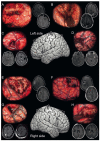
Figure 1
Figure 1
Illustrative cases. Illustrative cases of isocitrate dehydrogenase (IDH)-wildtype glioblastoma awake resection using direct cortico-subcortical electric stimulations to define functional boundaries. Intraoperative photographs showing the surgical field with the functional boundaries of the resection marked intraoperatively with numbered tags in the surgical cavity and corresponding pre- and postoperative magnetic resonance post-contrast T1-weighted imaging. (A) A 46-year-old right-handed woman underwent a supratotal awake resection (36.9 cm3, no residual tumor) for a left frontal IDH-wildtype glioblastoma. (Mapping was performed at 2.0 mA. Numbered tags: involuntary movement of the mouth and tongue at 1 and 2, involuntary movement of the hand at 3 and involuntary movement of the elbow at 4 identifying the primary motor cortical pathways; paresthesias of the tongue at 20 identifying the sensory cortical pathways; anarthria at 10 and 11 identifying language cortical pathways; arrest of voluntary movements of the upper limb at 5 and 6 and arrest of voluntary movements of the upper limb and of speech at 7 identifying cortico-subcortical negative motor networks; phonemic paraphasias at 12, 14, and 15 identifying the language subcortical dorsal phonologic pathway). (B) A 45-year-old right-handed man underwent a partial awake resection (62.5 cm3, 19.7 cm3 of residual tumor) for a left parietal IDH-wildtype glioblastoma. (Mapping was performed at 5.0 mA. Numbered tags: involuntary movement of the hand occurred at 1 and 2 identifying the primary motor cortical pathways; paresthesias of the hand at 20 and 22 and paresthesias of the shoulder at 23 and 24 identifying the sensory cortical pathways; paresthesias of the lower back at 25 and 27 and paresthesias of the lower limb at 26 and 28 identifying the sensory subcortical pathways). (C) A 63-year-old right-handed woman underwent a total awake resection (14.6 cm3, no residual tumor) for a left frontal IDH-wildtype glioblastoma. (Mapping was performed at 5.0 mA. Numbered tags: anarthria occurred at 10 identifying language cortical pathways; dysarthria at 11 identifying the primary motor cortical pathways; phonemic paraphasias at 20 and 21 identifying the language subcortical dorsal phonologic pathway). (D) A 66-year-old right-handed woman underwent a total awake resection (19.6 cm3, no residual tumor) for a left temporal IDH-wildtype glioblastoma. (Mapping was performed at 5.0 mA. Numbered tags: anarthria occurred at 10, and semantic paraphasias at 13 and 14 identifying language cortical pathways; dysarthria at 11 and 12 identifying the primary motor cortical pathways; latency at 15 and 17 and semantic paraphasia at 16 identifying the language subcortical ventral semantic pathway). (E) A 44-year-old right-handed woman underwent a subtotal awake resection (18.9 cm3, 0.6 cm3 of residual tumor) for a right frontal IDH-wildtype glioblastoma. (Mapping was performed at 3.5 mA. Numbered tags: arrest of voluntary movements of the upper limb occurred at 2, 3 and 4 identifying cortico-subcortical negative motor networks; involuntary movements of the tongue at 5, 6 and 7 identifying subcortical primary motor pathways). (F) A 26-year-old left-handed man underwent a supratotal awake resection (37.9 cm3, no residual tumor) for a right frontal IDH-wildtype glioblastoma. (Mapping was performed at 2.0 mA. Numbered tags: involuntary movement of the jaw and tongue occurred at 1, 2 and 3, involuntary movement of the hand at 4, and involuntary movement of the wrist at 5 identifying the primary motor cortical pathways; paresthesias of the lips at 20 and 21, paresthesias of the thumb at 22, paresthesias of third, fourth, fifth fingers at 23 identifying the sensory cortical pathways; involuntary movements of the jaw at 7 identifying subcortical primary motor pathways; arrest of voluntary movements of the mouth at 6 and saccadic lateral deviation of the eyes at 8 identifying cortico-subcortical negative motor networks). (G) A 54-year-old left-handed man underwent a total awake resection (2.4 cm3, no residual tumor) for a right parietal IDH-wildtype glioblastoma. (Mapping was performed at 3.0 mA. Numbered tags: involuntary movement of the wrist occurred at 1 and 2 identifying the primary motor cortical pathways; paresthesias of thumb at 20, paresthesias of second and third fingers at 24, paresthesias of fourth and fifth fingers at 21 and 23 identifying the sensory cortical pathways; latency during picture naming at 15; paresthesias of the thorax at 25, paresthesias of the upper limb at 27, and paresthesias of the lower limb at 27 identifying the sensory subcortical pathways). (H) A 54-year-old right-handed woman underwent a supratotal awake resection (28.9 cm3, no residual tumor) for a right frontal IDH-wildtype glioblastoma. (Mapping was performed at 3.5 mA. Numbered tags: dysarthria occurred at 10 identifying the primary motor cortical pathways; involuntary movements of the jaw at 2 and of the lips at 3 identifying subcortical primary motor pathways).
Figure 2
Figure 2
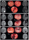
Illustrative cases of the intraoperative management of tumor-related elevated intracranial pressure and local mass effect during awake resection using intraoperative direct cortico-subcortical electrostimulation mapping of IDH-wildtype glioblastomas. On the left, preoperative magnetic resonance examinations (post-contrast T1-weighted and Fluid Attenuated Inversion Recovery sequences); in the middle, intraoperative photographs with eloquent sites tagged; on the right, day-one postoperative magnetic resonance examinations (post-contrast T1-weighted and Fluid Attenuated Inversion Recovery sequences). (A) A 63-year-old right-handed man presented with uncontrolled focal seizures revealing a left cystic contrast-enhancing necrotic parietal tumor. An awake resection was performed using intraoperative direct cortico-subcortical electrostimulation mapping. The initial cortical mapping (up to 6.0 mA) failed at identifying any eloquent sites. Intralesional debulking was performed, which reduced mass effect. Subsequent cortical mapping allowed for the identification of the primary motor cortex of the face (1) and hand (2), and of latency and semantic paraphasia in the supramarginal gyrus (10, 11, 12, 13). Then, the resection was performed beyond the limits of the solid tumor tissue component according to subcortical functional boundaries, with the arcuate fasciculus as the lateral limit of the surgical cavity (latency, 14, 15, 16). No visual disturbances were observed at the inferior limits of the surgical cavity. (B) A 39-year-old right-handed woman presented with signs of elevated intracranial pressure and language impairment revealing a left cystic contrast-enhancing necrotic frontal tumor. An awake resection was performed using intraoperative direct cortico-subcortical electrostimulation mapping. The initial cortical mapping (3.5 mA) allowed for the identification of the ventral premotor cortex inducing speech arrest (10, 11) and of the primary motor cortex of the hand (1, 2) with no other response elicited on cortical mapping. An ultrasound-guided cyst puncture was performed to reduce mass effect, which revealed the cortical negative motor networks inducing arrest of voluntary movements of the hand (3, 4) upon electrostimulation. Then, the resection was performed beyond the limits of the solid tumor tissue component according to subcortical functional boundaries, with the white matter involved in motor control as the posterior limit of the surgical cavity (arrest of voluntary movements, 5, 6, 7) and the arcuate fasciculus as the lateral limit of the surgical cavity (phonemic paraphasia, 12). (C) A 43-year-old right-handed man presented with focal seizures revealing a left contrast-enhancing and necrotic parietal tumor. An awake resection was performed using intraoperative direct cortico-subcortical electrostimulation mapping. Upon opening the dura, brain herniation occurred, and the patient experienced headaches, leading to initial cortical mapping failure. Intralesional debulking was performed, which reduced the mass effect and headaches. Subsequent cortical mapping (4.0 mA) allowed for the identification of the primary sensory cortex of the hand (1) and upper limb (4) and of semantic paraphasia in the supramarginal gyrus (10). Then, the resection was performed beyond the limits of the solid tumor tissue component according to subcortical functional boundaries, with the white matter involved in sensory control as the anterior limit of the surgical cavity (11, 12, 13, 14 for the lower limb; 15 for the upper limb). No visual or language disturbances were observed at the lateral and inferior limits of the surgical cavity.
Figure 3
Figure 3
Kaplan–Meier estimates of progression-free and overall survival. (A) Kaplan–Meier estimates of progression-free survival (left) and overall survival (right) in the whole study population (434 patients). (B) Kaplan–Meier estimates of progression-free survival (left) and overall survival (right) according to the first-line oncological treatment received following surgery: standard radiochemotherapy (Stupp, blue line), other oncological treatment (Other, green line), and no treatment (No treatment, red line). (C) Kaplan–Meier estimates of progression-free survival (left) and overall survival (right) according to the type of surgery performed: biopsy (Biopsy, red line), surgical resection under asleep conditions (Asleep, green line), and surgical resection under awake conditions (awake, blue line). (D) Kaplan–Meier estimates of progression-free survival (left) and overall survival (right) according to the extent of surgical resection: biopsy (Biopsy, red line), partial surgical resection (Partial, green line), total surgical resection (total, light blue line), and supratotal surgical resection (supratotal, blue line).

Similar articles

Cited by

References

    1. Stupp R., Mason W.P., van den Bent M.J., Weller M., Fisher B., Taphoorn M.J., Belanger K., Brandes A.A., Marosi C., Bogdahn U., et al. Radiotherapy plus concomitant and adjuvant temozolomide for glioblastoma. N. Engl. J. Med. 2005;352:987–996. doi: 10.1056/NEJMoa043330. - DOI - PubMed
    1. Hegi M.E., Diserens A.-C., Gorlia T., Hamou M.-F., de Tribolet N., Weller M. MGMT gene silencing and benefit from te-mozolomide in glioblastoma. N. Engl. J. Med. 2005;352:997–1003. doi: 10.1056/NEJMoa043331. - DOI - PubMed
    1. Molinaro A.M., Hervey-Jumper S., Morshed R.A., Young J., Han S.J., Chunduru P., Zhang Y., Phillips J.J., Shai A., Lafontaine M., et al. Association of maximal extent of resection of contrast-enhanced and non–contrast-enhanced tumor with survival within molecular subgroups of patients with newly diagnosed glioblastoma. JAMA Oncol. 2020;6:495–503. doi: 10.1001/jamaoncol.2019.6143. - DOI - PMC - PubMed
    1. Chambless L.B., Kistka H.M., Parker S.L., Hassam-Malani L., McGirt M.J., Thompson R.C., Mohammad L. The relative value of postoperative versus preoperative Karnofsky Performance Scale scores as a predictor of survival after surgical resection of glioblastoma multiforme. J. Neuro Oncol. 2014;121:359–364. doi: 10.1007/s11060-014-1640-x. - DOI - PubMed
    1. Sanai N., Polley M.Y., McDermott M.W., Parsa A.T., Berger M.S. An extent of resection threshold for newly diagnosed glioblastomas. J. Neurosurg. 2011;115:3–8. doi: 10.3171/2011.2.JNS10998. - DOI - PubMed