. 2021 Jun 8;10(6):1427.
doi: 10.3390/cells10061427.

Inhibition of Human Malignant Pleural Mesothelioma Growth by Mesenchymal Stromal Cells

Affiliations

Inhibition of Human Malignant Pleural Mesothelioma Growth by Mesenchymal Stromal Cells

Valentina Coccè et al. Cells. .

Abstract

Background: Malignant Pleural Mesothelioma (MPM) is an aggressive tumor that has a significant incidence related to asbestos exposure with no effective therapy and poor prognosis. The role of mesenchymal stromal cells (MSCs) in cancer is controversial due to their opposite effects on tumor growth and in particular, only a few data are reported on MSCs and MPM.

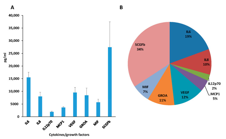
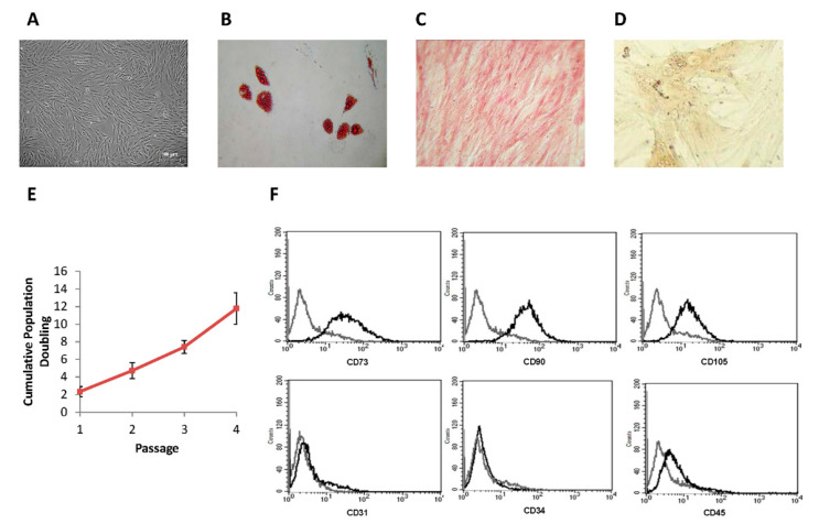
Methods: We investigated the in vitro efficacy of adipose tissue-derived MSCs, their lysates and secretome against different MPM cell lines. After large-scale production of MSCs in a bioreactor, their efficacy was also evaluated on a human MPM xenograft in mice.

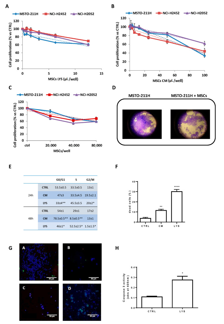
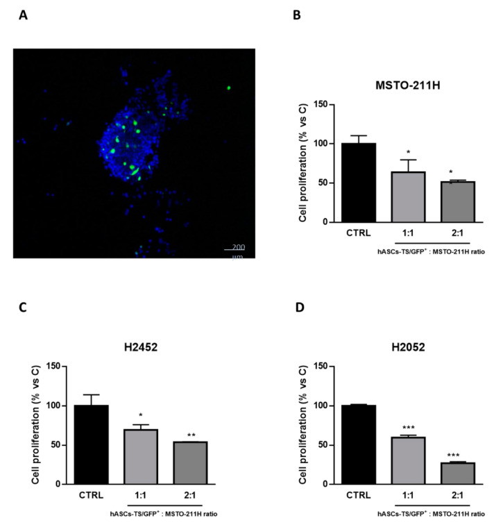
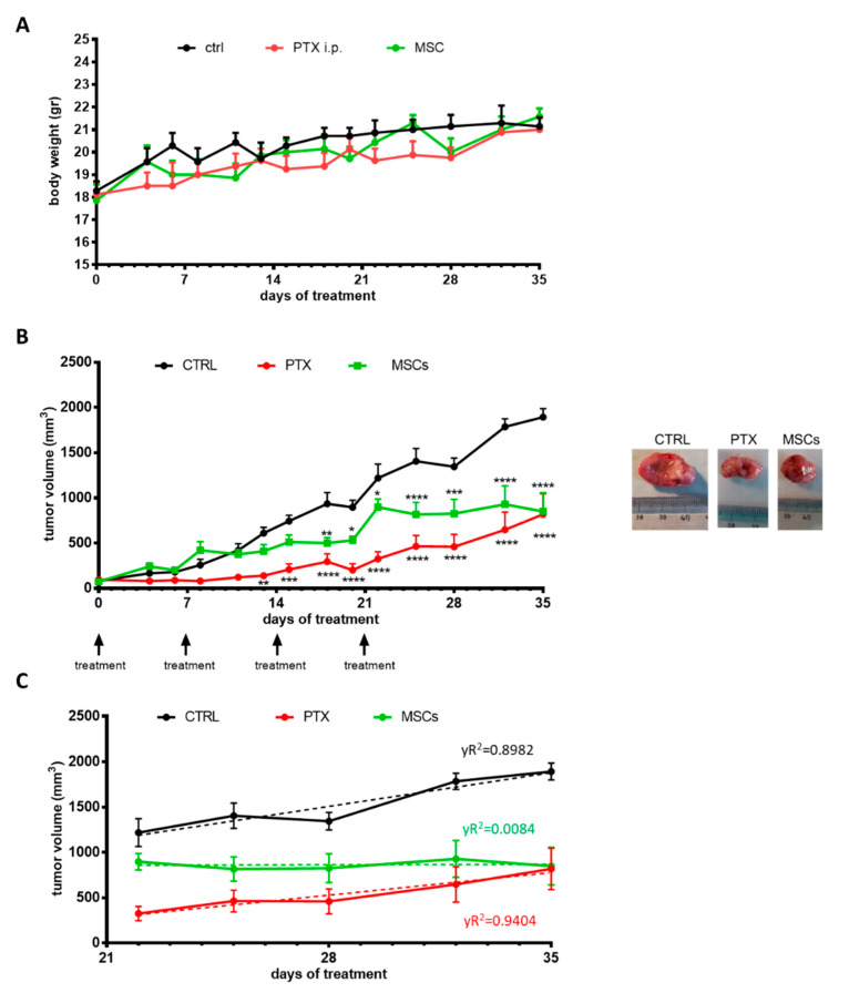
Results: MSCs, their lysate and secretome inhibited MPM cell proliferation in vitro with S or G0/G1 arrest of the cell cycle, respectively. MSC lysate induced cell death by apoptosis. The efficacy of MSC was confirmed in vivo by a significant inhibition of tumor growth, similar to that produced by systemic administration of paclitaxel. Interestingly, no tumor progression was observed after the last MSC treatment, while tumors started to grow again after stopping chemotherapeutic treatment.

Conclusions: These data demonstrated for the first time that MSCs, both through paracrine and cell-to-cell interaction mechanisms, induced a significant inhibition of human mesothelioma growth. Since the prognosis for MPM patients is poor and the options of care are limited to chemotherapy, MSCs could provide a potential new therapeutic approach for this malignancy.

Keywords: cell therapy; malignant pleural mesothelioma (MPM); mesenchymal stromal cells; mesothelioma.


Conflict of interest statement

The authors declare no conflict of interest.

Figures

Figure 1
Figure 1
Inhibitory effect of MSCs LYS and MSCs CM on mesothelioma proliferation and co-culture assay. The activity on cell proliferation of MSCs LYS (A) and MSCs CM (B), expressed as µL/well from 3 donors of adipose tissue-derived MSCs, were evaluated after 7 days in MSTO-211H, NCI-H2452 and NCI-H2052 cell lines by an MTT assay. Each point represents the mean ± standard error (SEM) of three replicates. Linear regression was reported (dashed lines). (C) MSTO-211H, NCI-H2452 and NCI-H2052 cells were co-cultured with MSCs and after 5 days, cell proliferation was evaluated by a crystal violet assay. Data are means ± SEM of six independent replicates. Linear regression was reported (dashed line). (D) Representative images of transwell inserts with MSTO-211H cells co-cultured alone or in the presence of 8 × 10⁴ MSCs after staining with crystal violet (400×). (E) MSTO-211H cells were treated with MSCs LYS (1:4) and MSCs CM (1:2). After 24 and 48 h, cells were stained with propidium iodide and analyzed by flow cytometry for cell cycle phase distribution. Percentage values ± SEM of two independent experiments are reported in the table (* p < 0.05, ** p < 0.01 vs. control). (F) MSTO-211H cells were treated with MSCs LYS (1:4) and MSCs CM (1:2). After 72 h, cell death was quantified by fluorescence microscopy analysis on Hoechst 33342 and propidium iodide-stained cells. Data are expressed as percentage values ± SEM of three independent experiments (** p < 0.01, **** p < 0.0001 vs. control). (G) Representative confocal images of control cells (A,C) and cells treated for 48 h with MSCs LYS 1:2 (B,D). Healthy viable cells were stained with CytoCalcein Violet 450 (blue), necrotic cells with 7-aminoactinomycin D (red), and apoptotic cells with phosphatidylserine (green). (A,B): adherent cells; (C,D): detached cells. (Objective 100×). (H) Caspase-3 activity measurement after 48 h of treatment. The histogram represents absorbance (abs) at 405 nm. Data are expressed as means ± SEM of three replicates. * p<0.05 vs. control.
Figure 2
Figure 2
Interaction of MSCs with MSTO-211H cells in vitro. (A) The cell-to-cell interference/interaction between MSCs and tumor cells was evaluated by mixing fluorescent MSCs (hASCs-TS/GFP+) with MSTO-211H cells and further analyzing the cyto-inclusion under a confocal microscope. (B) MSTO-211H, NCI-H2452 and NCI-H2052 cells were co-cultured in absence (ctrl) or in presence of hASCs-TS/GFP+ cells in the ratio of 1:1 or 1:2. After 2 days for MSTO-211H and after 4 days for NCI-H2452 and NCI-H2052, the cells were detached and counted under a fluorescence microscopy. Data are the means ± SEM of 3 replicates. * p < 0.05, ** p < 0.01, *** p < 0.001 vs. control.
Figure 3
Figure 3
MSCs secretome analysis. (A) The histogram reports eight cytokines/growth factors measured in MSC-conditioned media (expressed in pg/mL). Each point represents the mean ± standard error (SEM) of the determinations performed on nine different MSCs donors. (B) Cytokines/growth factors expressed as percentage calculated on the total amount of the eight molecules.
Figure 4
Figure 4
MSC characterization. (A): Spindle-shaped MSCs morphology (magnification 5×). (B): Adipogenic differentiation evaluated by Oil Red staining (presence of red cytoplasmic inclusions); (C): Osteogenic differentiation evaluated by Alizarin Red S staining; (D): Chondrogenic differentiation evaluated by micro-mass methods. (All at 200× magnification). (E): Number of cumulative population doubling with respect to cellular passage. Each point represents the mean ± SEM of 14 replicates. (F): Phenotypic characterization by FACS of a representative sample of MSCs.
Figure 5
Figure 5
Effects of MSCs in MSTO-211H xenograft model. (A) MSTO-211H cells were subcutaneously inoculated into BALB/C nude mice, and after tumors had reached an average size of approximately 100 mm3, the animals were treated once a week (at days 0, 7, 14 and 21) with vehicle alone (CTRL), paclitaxel (20 mg/kg) or MSCs (5 × 10⁶). (A) Mice body weight was monitored for the entire duration of the treatment. (B) Tumor volumes were measured twice per week and data are expressed as means of ± SEM. * p < 0.05, ** p < 0.01, *** p < 0.001, **** p < 0.0001 vs. CTRL. Representative images of dissected xenograft tumors are shown. (C) The graph shows the tumor growth during the 14 days after stopping treatments (from 22 to 35 days). Data are expressed as means ± SEM and for each group, the linear regression (dashed line) with R2 value is reported.
Figure 6
Figure 6
Morphometric analysis of tumor xenografts. Selected sections from MSTO-211H xenografts untreated (CTRL, A), treated with paclitaxel (PTX) (B) or with MSCs (C) stained by Masson’s Trichrome to distinguish the fibrotic tissue (greenish) from neoplastic cells (purple). Black arrows in B and C point to necrotic areas, some of which show pigmented (reddish) debris. Red asterisks in A and C indicate skin and adnexa. Scale bars = 100 µm. (D) Bar graph showing the quantitative evaluation of tissue composition (neoplastic tissue, fibrosis and necrosis) in tumor xenografts. Data are expressed as means ± SEM. * p < 0.05, ** p < 0.02 vs. control group.

Similar articles

Cited by

References

    1. Carbone M., Ly B.H., Dodson R.F., Pagano I., Morris P.T., Dogan U.A., Gazdar A.F., Pass H.I., Yang H. Malignant mesothelioma: Facts, myths, and hypotheses. J. Cell Physiol. 2012;227:44–58. doi: 10.1002/jcp.22724. - DOI - PMC - PubMed
    1. Peto J., Decarli A., La Vecchia C., Levi F., Negri E. The European mesothelioma epidemic. Br. J. Cancer. 1999;79:666–672. doi: 10.1038/sj.bjc.6690105. - DOI - PMC - PubMed
    1. Fennell D.A., Gaudino G., O’Byrne K.J., Mutti L., van Meerbeeck J. Advances in the systemic therapy of malignant pleural mesothelioma. Nat. Clin. Pract. Oncol. 2008;5:136–147. doi: 10.1038/ncponc1039. - DOI - PubMed
    1. Krug L.M., Pass H.I., Rusch V.W., Kindler H.L., Sugarbaker D.J., Rosenzweig K.E., Flores R., Friedberg J.S., Pisters K., Monberg M., et al. Multicenter phase II trial of neoadjuvant pemetrexed plus cisplatin followed by extrapleural pneumonectomy and radiation for malignant pleural mesothelioma. J. Clin. Oncol. 2009;27:3007–3013. doi: 10.1200/JCO.2008.20.3943. - DOI - PMC - PubMed
    1. Mutti L., Peikert T., Robinson B.W.S., Scherpereel A., Tsao A.S., de Perrot M., Woodard G.A., Jablons D.M., Wiens J., Hirsch F.R., et al. Scientific Advances and New Frontiers in Mesothelioma Therapeutics. J. Thorac. Oncol. 2018;13:1269–1283. doi: 10.1016/j.jtho.2018.06.011. - DOI - PMC - PubMed

Publication types