Review
. 2021 Jun 8;10(6):1428.
doi: 10.3390/cells10061428.

Primary Cilium Is Involved in Stem Cell Differentiation and Renewal through the Regulation of Multiple Signaling Pathways

Affiliations
Review

Primary Cilium Is Involved in Stem Cell Differentiation and Renewal through the Regulation of Multiple Signaling Pathways

Sila Yanardag et al. Cells. .

Abstract

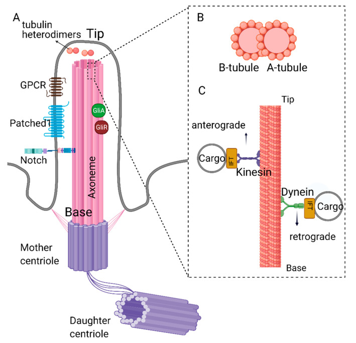
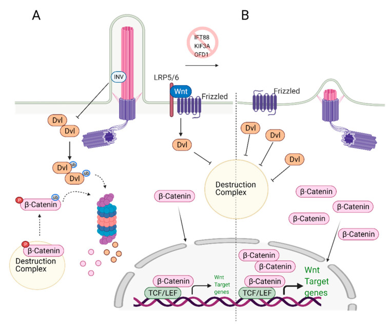
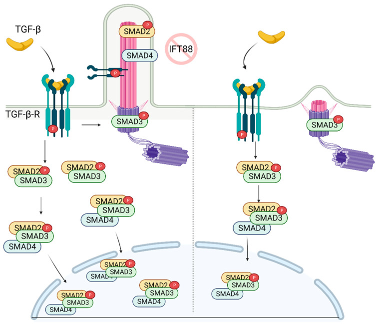
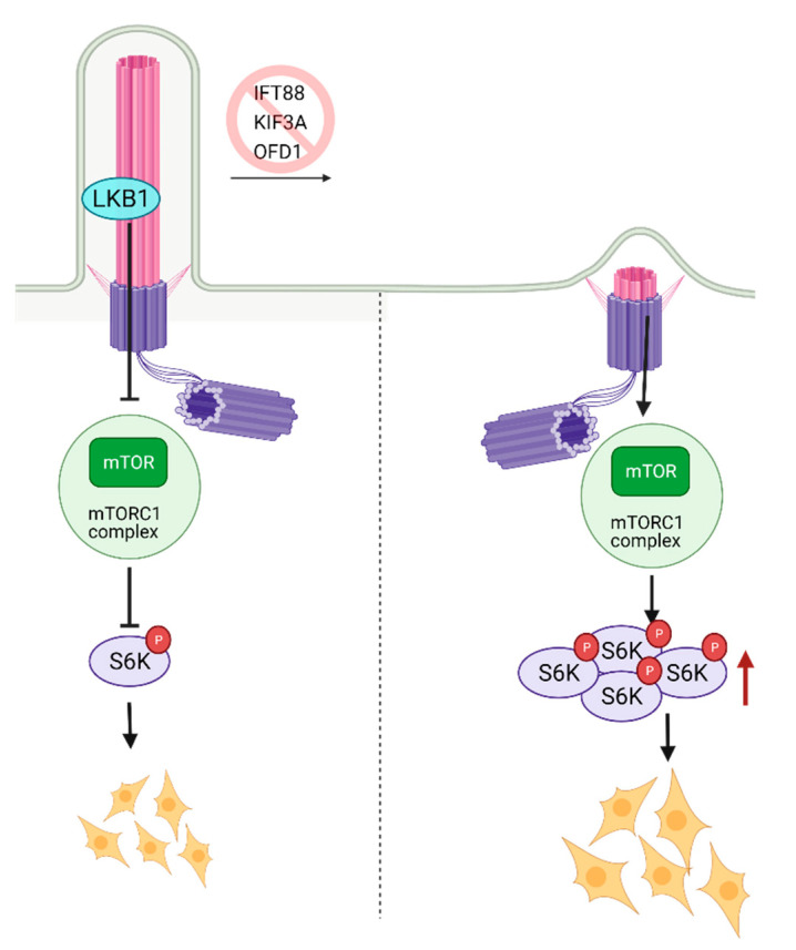
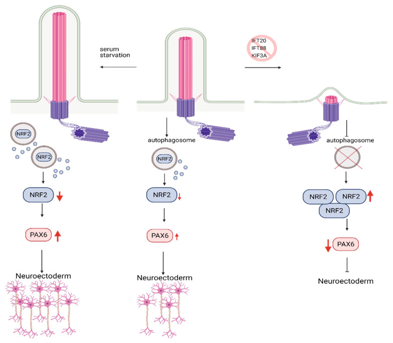
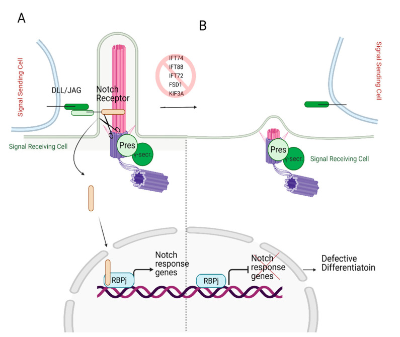
Signaling networks guide stem cells during their lineage specification and terminal differentiation. Primary cilium, an antenna-like protrusion, directly or indirectly plays a significant role in this guidance. All stem cells characterized so far have primary cilia. They serve as entry- or check-points for various signaling events by controlling the signal transduction and stability. Thus, defects in the primary cilia formation or dynamics cause developmental and health problems, including but not limited to obesity, cardiovascular and renal anomalies, hearing and vision loss, and even cancers. In this review, we focus on the recent findings of how primary cilium controls various signaling pathways during stem cell differentiation and identify potential gaps in the field for future research.

Keywords: Notch; TGF; Wnt; cancer stem cells; differentiation; mTOR; primary cilia; signaling; stem cells.


Conflict of interest statement

The authors declare no conflict of interest.

Figures

Figure 1
Figure 1
Structure of primary cilia. (A) Primary cilium is nucleated from the mother centriole of the basal body. Axoneme consists of 9 microtubule doublets formed by α and β tubulin heterodimers. (B) Cross-section of complete A- and partial B-tubules. (C) Dynein and kinesin proteins carrying cargo on the axoneme. Retrograde movement (from tip to the base) is carried out by dynein proteins, whereas kinesin proteins carry out anterograde movement (from base to the tip). Kinesin and dynein proteins are essential for the assembly and disassembly of the primary cilium as they carry building blocks or the depolymerizing agents along the axoneme. Created with BioRender.com.
Figure 2
Figure 2
Role of primary cilia in Wnt/β-catenin regulation and stem cell biology. (A) Primary cilium keeps the expression of Wnt target genes in check. Upon binding of Wnt ligands to the receptors, Disheveled (Dvl) inactivates the destruction complex, causing release and translocation of β-catenin to the nucleus. In the nucleus, it serves as a transcription activator and initiates the expression of Wnt target genes. At the same time, excess Dvl is targeted to proteasomal degradation via ciliary-localized Inversin (INV), causing degradation of some of the β-catenin. (B) In the absence of primary cilium, increased cytoplasmic localization of Dvl is observed. This causes increased levels of cytoplasmic and nuclear b-catenin, leading to overexpression of Wnt/β-catenin target genes. Created with BioRender.com.
Figure 3
Figure 3
TGF-β signaling is regulated by primary cilia. In the presence of primary cilia, p-TGF-β-RI, p-SMAD2, and SMAD4 are localized on the cilia, while p-SMAD3 is localized on the ciliary base. Stimulation of the receptor with TGF-β ligand results in increased nuclear localization of active SMAD2/3/4 complex. In the absence of cilia, while p-SMAD3 is still localized on the ciliary base, its nuclear localization is diminished as well as the abundance of SMAD2/3 complex in the cell. Created with BioRender.com.
Figure 4
Figure 4
Regulation of mTOR signaling through primary cilia. Ciliary localized LKB1 inhibits mTOR, and keeps mTORC1 activity at a lower level. Inhibition of primary cilia formation results in increased mTORC1 activity, increased levels of active S6K, and larger cell size. Created with BioRender.com.
Figure 5
Figure 5
Role of Autophagy and primary cilia in embryonic stem cell lineage specification. Primary cilium promotes autophagosome formation in hESCs, keeping embryonic development in check via the controlled degradation of Nrf2. Inhibition of primary cilia formation results in increased levels of Nrf2 due to constrained autophagosome formation. Increased levels of Nrf2 reduce the expression of PAX6, neuroectoderm fate-determinant transcription factor, which in turn results in inhibition of hESC’s commitment to neuroectoderm lineage. Conversely, activation of primary cilia via serum starvation causes higher levels of PAX6 expression and promotes commitment to neuroectoderm lineage. Created with BioRender.com.
Figure 6
Figure 6
Role of primary cilia in Notch signaling. (A) Notch receptor localizes on the primary cilium. Following the binding of the Notch receptor to the ligands DLL/JAG, NICD is cleaved by presenilin (Pres), the catalytic subunit of γ-secretase, which localizes on the basal body. Cleaved NICD translocates to the nucleus. It serves as a transcriptional co-activator of the RBPj (Recombination signal Binding Protein for immunoglobulin kappa j region) to express Notch response genes. (B) In the absence of primary cilia, expression of Notch target genes is halted since Notch receptor cannot localize to the ciliary membrane and be cleaved by presenilin, resulting in defective differentiation. Created with BioRender.com.

Similar articles

Cited by

References

    1. Loskutov Y.V., Griffin C.L., Marinak K.M., Bobko A., Margaryan N.V., Geldenhuys W.J., Sarkaria J.N., Pugacheva E.N. LPA signaling is regulated through the primary cilium: A novel target in glioblastoma. Oncogene. 2018;37:1457–1471. doi: 10.1038/s41388-017-0049-3. - DOI - PMC - PubMed
    1. Sun S., Fisher R.L., Bowser S.S., Pentecost B.T., Sui H. Three-dimensional architecture of epithelial primary cilia. Proc. Natl. Acad. Sci. USA. 2019;116:9370–9379. doi: 10.1073/pnas.1821064116. - DOI - PMC - PubMed
    1. Plotnikova O.V., Golemis E.A., Pugacheva E.N. Cell cycle-dependent ciliogenesis and cancer. Cancer Res. 2008;68:2058–2061. doi: 10.1158/0008-5472.CAN-07-5838. - DOI - PMC - PubMed
    1. Allen R.D. A reinvestigation of cross-sections of cilia. J. Cell Biol. 1968;37:825–831. doi: 10.1083/jcb.37.3.825. - DOI - PMC - PubMed
    1. Stepanek L., Pigino G. Microtubule doublets are double-track railways for intraflagellar transport trains. Science. 2016;352:721–724. doi: 10.1126/science.aaf4594. - DOI - PubMed

Publication types