. 2021 Jun 8;13(12):1903.
doi: 10.3390/polym13121903.

Influence of ER-CR-YSGG Laser and Photodynamic Therapy on the Dentin Bond Integrity of Nano-Hydroxyapatite Containing Resin Dentin Adhesive: SEM-EDX, Micro-Raman, Micro-Tensile, and FTIR Evaluation

Affiliations

Influence of ER-CR-YSGG Laser and Photodynamic Therapy on the Dentin Bond Integrity of Nano-Hydroxyapatite Containing Resin Dentin Adhesive: SEM-EDX, Micro-Raman, Micro-Tensile, and FTIR Evaluation

Abdullah S Aljamhan et al. Polymers (Basel). .

Abstract

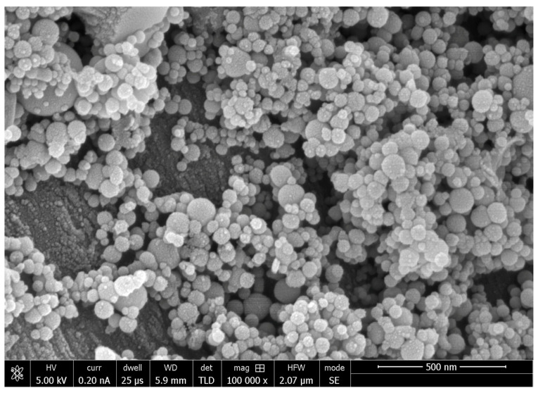
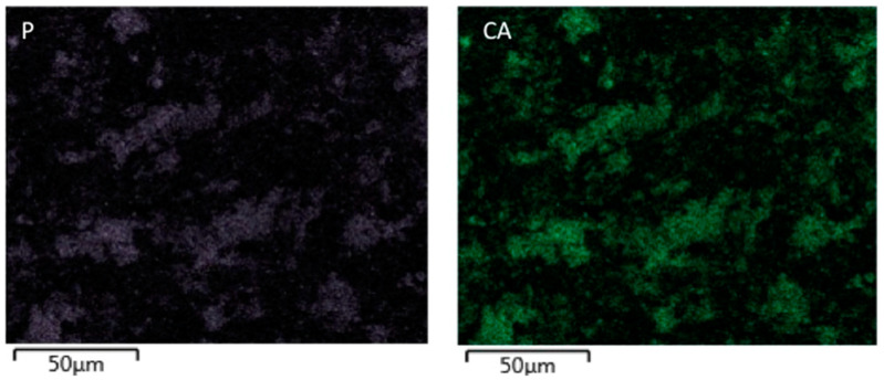
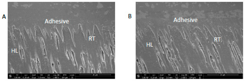
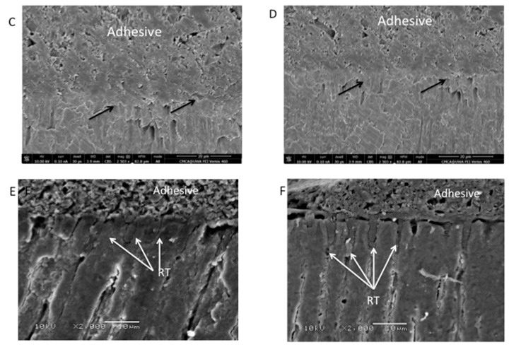
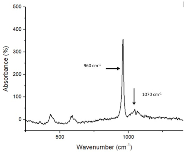
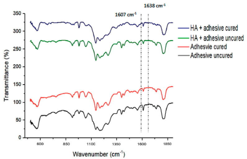
The study aimed to analyze the effect of the addition of nano-hydroxyapatite (nano-HA) particles on the mechanical properties of experimental adhesive (EA). Furthermore, dentin interaction of EA (without nano-HA) and EA with nano-HA (hereon referred to as HA-10%) were also investigated and equated. Methods consisting of scanning electron microscopy (SEM)-energy-dispersive X-ray spectroscopy (EDX), micro-Raman spectroscopy, micro-tensile bond strength (µTBS) test, and Fourier transform infrared (FTIR) spectroscopy were employed to study nano-HA particles shape, dentin bond strength, degree of conversion (DC), and adhesive-dentin interaction. Ninety teeth (N = 90) were collected, and pre-bonding, conditioning of dentin was performed utilizing phosphoric acid (H3PO4) etching, photodynamic therapy (PDT), and ER-CR-YSGG (ECY) laser. The teeth were set to form bonded specimens using two adhesives. Nano-HA particles were spherical-shaped, and EDX confirmed the presence of oxygen, calcium, and phosphorus. Micro-Raman spectroscopy revealed distinct phosphate and carbonate peaks for nano-HA. The µTBS test demonstrated highest values for HA-10% group on the H3PO4 conditioned dentin. The greatest DC was observed for the EA group. The addition of nano-HA-10 wt.% particles in dentin adhesive resulted in improved bond strength. The incorporation also demonstrated acceptable DC (although lower than EA group), suitable dentin interaction, and resin tag formation.

Keywords: H3PO4; adhesive; hydroxyapatite; laser; photodynamic therapy.


Conflict of interest statement

The authors declare no conflict of interest.

Figures

Figure 1
Figure 1
SEM image of HA nanoparticles. These nanoparticles on SEM showed a uniform round shape in mostly agglomerated form with few isolated particles.
Figure 2
Figure 2
EDX element mapping evaluation of the HA nanoparticles. The two figures show evidence of calcium (CA), and phosphorus (P) in the samples.
Figure 3
Figure 3
SEM images of bonded dentin specimen. (A) HA-PDT group specimens showing standard hybrid layer (HL) formation with low to average resin tag formation in dentin. (B) HA-ER group specimens showing classic HL formation with a high number of resin tags. (C) HA-ECY group specimens showing uneven dentin surface and craters (arrows) at the interface with no resin tags and dentin penetration. (D) EA-ECY group specimens displaying craters (arrows) on the dentin surface at the interface with little to no HL and missing resin tags, showing minimal dentin penetration. (E) EA-PDT group specimens showing a low number of resin tags in dentin. (F) EA-ER group specimens showing standard HL and multiple resin tag formation.
Figure 3
Figure 3
SEM images of bonded dentin specimen. (A) HA-PDT group specimens showing standard hybrid layer (HL) formation with low to average resin tag formation in dentin. (B) HA-ER group specimens showing classic HL formation with a high number of resin tags. (C) HA-ECY group specimens showing uneven dentin surface and craters (arrows) at the interface with no resin tags and dentin penetration. (D) EA-ECY group specimens displaying craters (arrows) on the dentin surface at the interface with little to no HL and missing resin tags, showing minimal dentin penetration. (E) EA-PDT group specimens showing a low number of resin tags in dentin. (F) EA-ER group specimens showing standard HL and multiple resin tag formation.
Figure 4
Figure 4
Raman spectra of the HA nanoparticles showing chemical groups identified by Raman analysis that could be identified as phosphate (υ1 PO4 ≈ 960 cm−1) and carbonate group at 1070 cm−1.
Figure 5
Figure 5
CH3 and –CH2 deformations within the EA are represented by peaks between 1410 and 1460 cm−1.
Figure 6
Figure 6
FTIR spectrum of uncured and cured resin adhesives containing HA nanoparticles and control (without nanoparticles). The DC was estimated from 1607 cm−1 and 1638 cm−1 wavenumber.

Similar articles

Cited by

References

    1. Tsujimoto A., Barkmeier W.W., Fischer N.G., Nojiri K., Nagura Y., Takamizawa T., Latta M.A., Miazaki M. Wear of resin composites: Current insights into underlying mechanisms, evaluation methods and influential factors. Jpn. Dent. Sci. Rev. 2018;54:76–87. doi: 10.1016/j.jdsr.2017.11.002. - DOI - PMC - PubMed
    1. Opdam N.J., Bronkhorst E.M., Loomans B.A., Huysmans M.C. 12-year survival of composite vs. amalgam restorations. J. Dent. Res. 2010;89:1063–1067. doi: 10.1177/0022034510376071. - DOI - PubMed
    1. Bourbia M., Finer Y. Biochemical stability and interactions of dental resin composites and adhesives with host and bacteria in the oral cavity: A review. J. Can. Dent. Assoc. 2018;84:i1. - PubMed
    1. Leitune V.C., Collares F.M., Trommer R.M., Andrioli D.G., Bergmann C.P., Samuel S.M. The addition of nanostructured hydroxyapatite to an experimental adhesive resin. J. Dent. 2013;41:321–327. doi: 10.1016/j.jdent.2013.01.001. - DOI - PubMed
    1. Sofan E., Sofan A., Palaia G., Tenore G., Romeo U., Migliau G. Classification review of dental adhesive systems: From the IV generation to the universal type. Ann. Stomatol. 2017;8:1–17. - PMC - PubMed

LinkOut - more resources