. 2021 Jun 8;12(6):883.
doi: 10.3390/genes12060883.

The Combined Human Genotype of Truncating TTN and RBM20 Mutations Is Associated with Severe and Early Onset of Dilated Cardiomyopathy

Affiliations

The Combined Human Genotype of Truncating TTN and RBM20 Mutations Is Associated with Severe and Early Onset of Dilated Cardiomyopathy

Anna Gaertner et al. Genes (Basel). .

Abstract

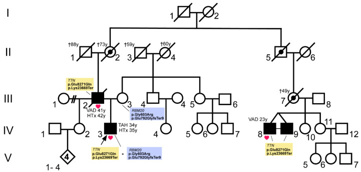
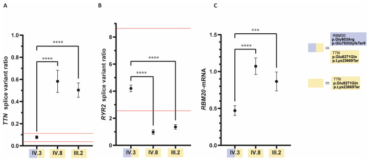
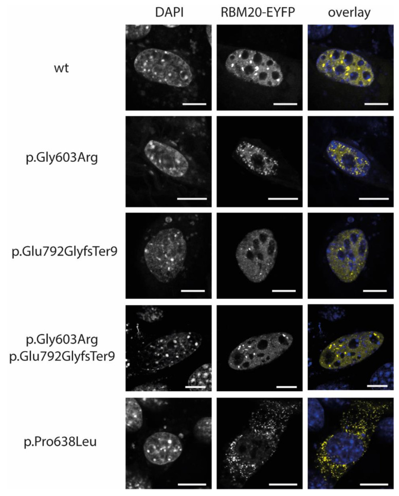
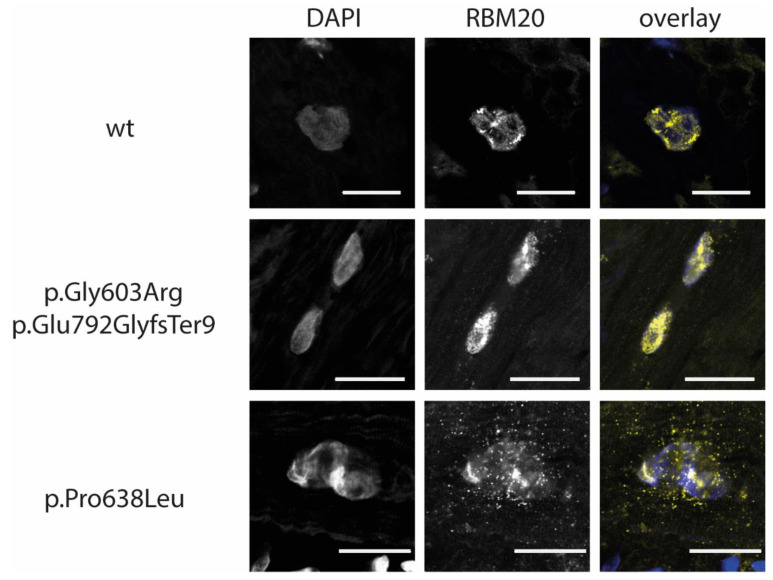
A major cause of heart failure is cardiomyopathies, with dilated cardiomyopathy (DCM) as the most common form. Over 40 genes are linked to DCM, among them TTN and RBM20. Next Generation Sequencing in clinical DCM cohorts revealed truncating variants in TTN (TTNtv), accounting for up to 25% of familial DCM cases. Mutations in the cardiac splicing factor RNA binding motif protein 20 (RBM20) are also known to be associated with severe cardiomyopathies. TTN is one of the major RBM20 splicing targets. Most of the pathogenic RBM20 mutations are localized in the highly conserved arginine serine rich domain (RS), leading to a cytoplasmic mislocalization of mutant RBM20. Here, we present a patient with an early onset DCM carrying a combination of (likely) pathogenic TTN and RBM20 mutations. We show that the splicing of RBM20 target genes is affected in the mutation carrier. Furthermore, we reveal RBM20 haploinsufficiency presumably caused by the frameshift mutation in RBM20.

Keywords: RBM20; TTN; cardiomyopathy; haploinsufficiency; mutation.


Conflict of interest statement

The authors declare that there is no conflict of interest.

Figures

Figure 1
Figure 1
Pedigree of a cardiomyopathy family with (likely) pathogenic RBM20 and TTN mutations. Circles represent females, squares males, slash denotes deceased. The index patient (IV.3) is marked with an arrow. Available myocardial tissue of patients with end-stage heart failure, who received heart transplantation (HTx) or mechanical circulatory support (left ventricular assist device (VAD) or total artificial heart (TAH)) were indicated with red heart symbols. Affected members with dilated cardiomyopathy (DCM) are shown in black and obligate mutation carriers of the TTN mutations are marked with a dot. The age of the patients in years (y) at the time of HTx, VAD- or TAH implantation or at the time of death (†) is specified. The respective genotypes of the analyzed patients are shown in the figure.
Figure 2
Figure 2
Quantitative real-time polymerase chain reaction (qRT-PCR) results of (A) TTN- (B) RYR2-splice variant ratios, and (C) relative quantification of RBM20 mRNA. Data from affected family members with available cardiac tissue were measured as technical quintuplicates and are shown as means with standard deviation (SD). For statistical analyses one-way analysis of variance (ANOVA) with Dunnett’s multiple comparisons test was used. **** = p < 0.0001, *** = p = 0.0001. (A,B) The TTN- and RYR2-splicing in the index patient (IV.3) is significantly different from the splicing in his affected relatives, who are only carriers of the TTN-mutations. The TTN- and RYR2-splicing of IV.3 are within the previously defined reference range [10] for pathogenic RBM20 mutations (red lines). (C) qRT-PCR analysis revealed decreased RBM20 mRNA expression in the RBM20 mutation carrier IV.3 in comparison to his relatives.
Figure 3
Figure 3
Localization analyses of RNA binding motif protein 20- enhanced yellow fluorescent protein (RBM20-EYFP) transfected C2C12 cells. RBM20 is shown in yellow, and the nuclei were labelled with 4’,6-diamidine-2’-phenylindole dihydrochloride (DAPI, blue) in the overlay. EYFP tagged wildtype RBM20 localizes in the nucleus. The mutant forms of RBM20 (p.Gly603Arg and p.Glu792GlyfsTer9) are comparably localized in the nuclei. As described previously [10], only the mutant form RBM20-p.Pro638Leu shows an abnormal cytoplasmic localization. Scale bars = 10 µm.
Figure 4
Figure 4
Immunohistochemical analysis of myocardial tissue of cardiomyopathy patients. Representative images of explanted myocardial tissue sections, which were labelled with primary anti RBM20 and Cy3-conjugated secondary antibodies (yellow in the overlay), are shown. Nuclei were labelled with DAPI (blue in the overlay). Tissue from a DCM patient with no RBM20 mutation was used as control. Comparable to the control sample RBM20 is localized in the nuclei in explanted myocardial tissue from IV.3. Sections from a DCM patient with the pathogenic RBM20 mutation p.Pro638Leu were used as a positive control for aberrant cytoplasmic RBM20 localization. Scale bars = 20 µm.
Figure 5
Figure 5
Integrated genome views of exon 8 of RBM20. The chromosomal position corresponding to RBM20 c.1807 is marked with an arrow. Shown are the aligned reads obtained by RNA-sequencing (A) or DNA-sequencing (B) in the respective chromosomal region of index patient IV.3 (RBM20 p.Gly603Arg+p.Glu792fsTer9 and TTN p.Glu8271Gln+ p.Lys23669Ter). At chromosomal position 10:110,810,389 approximately 15% of the RNA reads (A, bar on the left) represent the mutant form, whereas 46% of the reads (B, bar on the left) at chromosomal position 10:110,810,389 represent the mutant DNA form.

References

    1. Hershberger R.E., Hedges D.J., Morales A. Dilated cardiomyopathy: The complexity of a diverse genetic architecture. Nat. Rev. Cardiol. 2013;10:531–547. doi: 10.1038/nrcardio.2013.105. - DOI - PubMed
    1. Khush K.K., Cherikh W.S., Chambers D.C., Goldfarb S., Hayes D., Jr., Kucheryavaya A.Y., Levvey B.J., Meiser B., Rossano J.W., Stehlik J., et al. The International Thoracic Organ Transplant Registry of the International Society for Heart and Lung Transplantation: Thirty-fifth Adult Heart Transplantation Report-2018; Focus Theme: Multiorgan Transplantation. J. Heart Lung Transpl. 2018;37:1155–1168. doi: 10.1016/j.healun.2018.07.022. - DOI - PubMed
    1. Rossano J.W., Cherikh W.S., Chambers D.C., Goldfarb S., Hayes D., Jr., Khush K.K., Kucheryavaya A.Y., Toll A.E., Levvey B.J., Meiser B., et al. The International Thoracic Organ Transplant Registry of the International Society for Heart and Lung Transplantation: Twenty-first pediatric heart transplantation report-2018; Focus theme: Multiorgan Transplantation. J. Heart Lung Transpl. 2018;37:1184–1195. doi: 10.1016/j.healun.2018.07.018. - DOI - PubMed
    1. McMurray J.J., Adamopoulos S., Anker S.D., Auricchio A., Bohm M., Dickstein K., Falk V., Filippatos G., Fonseca C., Gomez-Sanchez M.A., et al. ESC Guidelines for the diagnosis and treatment of acute and chronic heart failure 2012: The Task Force for the Diagnosis and Treatment of Acute and Chronic Heart Failure 2012 of the European Society of Cardiology. Developed in collaboration with the Heart Failure Association (HFA) of the ESC. Eur. Heart J. 2012;33:1787–1847. doi: 10.1093/eurheartj/ehs104. - DOI - PubMed
    1. Brodehl A., Ebbinghaus H., Deutsch M.A., Gummert J., Gartner A., Ratnavadivel S., Milting H. Human Induced Pluripotent Stem-Cell-Derived Cardiomyocytes as Models for Genetic Cardiomyopathies. Int. J. Mol. Sci. 2019;20:4381. doi: 10.3390/ijms20184381. - DOI - PMC - PubMed

Publication types

Substances

LinkOut - more resources