

Diagnostic Value of Whole-Body MRI Short Protocols in Bone Lesion Detection in Multiple Myeloma Patients
- PMID: 34201122
- PMCID: PMC8226715
- DOI: 10.3390/diagnostics11061053
Diagnostic Value of Whole-Body MRI Short Protocols in Bone Lesion Detection in Multiple Myeloma Patients
Abstract
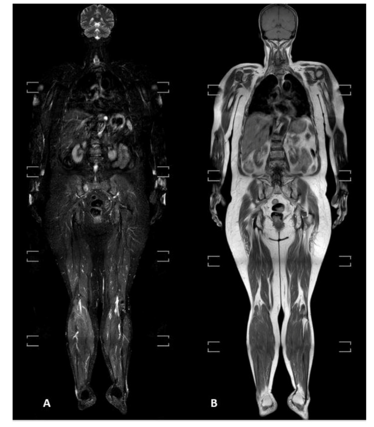
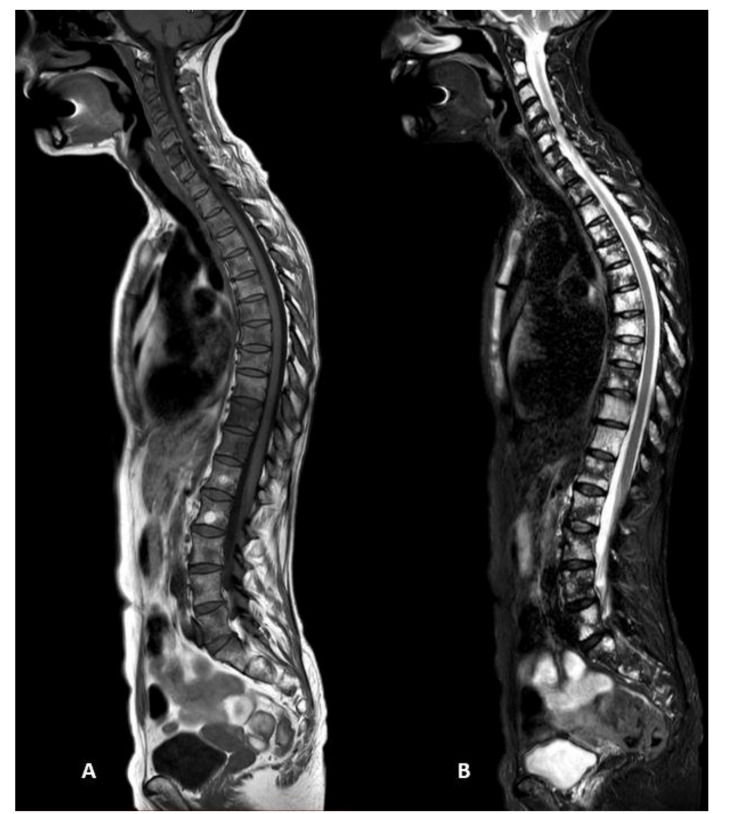
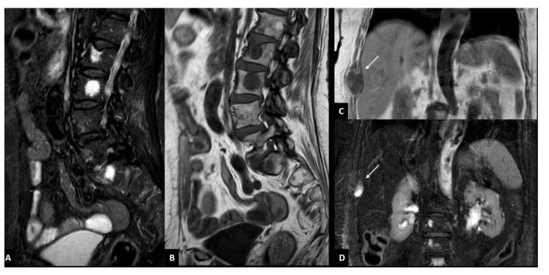
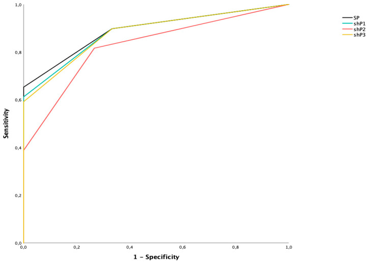
The aim of the study is to evaluate the effectiveness of short whole-body magnetic resonance imaging (WBMRI) protocols for the overall assessment of bone marrow involvement in patients with multiple myeloma (MM), in comparison with standard whole-body MRI protocol. Patients with biopsy-proven MM, who underwent a WBMRI with full-body coverage (from vertex to feet) were retrospectively enrolled. WBMRI images were independently evaluated by two expert radiologists, in terms of infiltration patterns (normal, focal, diffuse, and combined), according to location (the whole skeleton was divided into six anatomic districts: skull, spine, sternum and ribs, upper limbs, pelvis and proximal two-thirds of the femur, remaining parts of lower limbs) and lytic lesions number (<5, 5-20, and >20). The majority of patients showed focal and combined infiltration patterns with bone lesions predominantly distributed in the spine and pelvis. As skull and lower limbs are less frequently involved by focal bone lesions, excluding them from the standard MRI protocol allows to obtain a shorter protocol, maintaining a good diagnostic value.
Keywords: diffusion-weighted imaging; hematologic neoplasms; infiltration pattern; magnetic resonance imaging; multiple myeloma.
Conflict of interest statement
The authors declare no conflict of interest.
Figures
Similar articles
-
Whole Body Low Dose Computed Tomography (WBLDCT) Can Be Comparable to Whole-Body Magnetic Resonance Imaging (WBMRI) in the Assessment of Multiple Myeloma.Diagnostics (Basel). 2021 May 11;11(5):857. doi: 10.3390/diagnostics11050857. Diagnostics (Basel). 2021. PMID: 34064594 Free PMC article.
-
Prospective comparison of 18-FDG PET/CT and whole-body diffusion-weighted MRI in the assessment of multiple myeloma.Ann Hematol. 2020 Dec;99(12):2869-2880. doi: 10.1007/s00277-020-04265-2. Epub 2020 Sep 19. Ann Hematol. 2020. PMID: 32951093
-
18F-FDG PET/CT, 99mTc-MIBI, and MRI in evaluation of patients with multiple myeloma.J Nucl Med. 2008 Feb;49(2):195-200. doi: 10.2967/jnumed.107.045641. Epub 2008 Jan 16. J Nucl Med. 2008. PMID: 18199607
-
Whole-body magnetic resonance imaging (WBMRI) versus whole-body computed tomography (WBCT) for myeloma imaging and staging.Skeletal Radiol. 2022 Jan;51(1):43-58. doi: 10.1007/s00256-021-03799-4. Epub 2021 May 24. Skeletal Radiol. 2022. PMID: 34031705 Free PMC article. Review.
-
Role of MRI for the diagnosis and prognosis of multiple myeloma.Eur J Radiol. 2005 Jul;55(1):56-63. doi: 10.1016/j.ejrad.2005.01.017. Eur J Radiol. 2005. PMID: 15950101 Review.
Cited by
-
The importance of bone marrow infiltration patterns in multiple myeloma seen on magnetic resonance imaging-Case report and imaging perspective.Clin Case Rep. 2022 Oct 17;10(10):e6452. doi: 10.1002/ccr3.6452. eCollection 2022 Oct. Clin Case Rep. 2022. PMID: 36267826 Free PMC article.
References
-
- Rajkumar S.V., Dimopoulos M., Palumbo A., Blade J., Merlini G., Mateos M.-V., Kumar S., Hillengass J., Kastritis E., Richardson P., et al. International Myeloma Working Group updated criteria for the diagnosis of multiple myeloma. Lancet Oncol. 2014;15:e538–e548. doi: 10.1016/S1470-2045(14)70442-5. - DOI - PubMed
-
- Hillengass J., Usmani S., Rajkumar S.V., Durie B.G.M., Mateos M.V., Lonial S., Joao C., Anderson K.C., García-Sanz R., Riva E., et al. International myeloma working group consensus recommendations on imaging in monoclonal plasma cell disorders. Lancet Oncol. 2019;20:e302–e312. doi: 10.1016/S1470-2045(19)30309-2. - DOI - PubMed
-
- Landgren O., Kyle R.A., Pfeiffer R.M., Katzmann J.A., Caporaso N.E., Hayes R.B., Dispenzieri A., Kumar S., Clark R.J., Baris D., et al. Monoclonal gammopathy of undetermined significance (MGUS) consistently pre-cedes multiple myeloma: A prospective study. Blood. 2009;113:5412–5417. doi: 10.1182/blood-2008-12-194241. - DOI - PMC - PubMed
LinkOut - more resources
Full Text Sources
Research Materials
