. 2021 Jun 8;22(12):6185.
doi: 10.3390/ijms22126185.

T-Type Ca2+ Enhancer SAK3 Activates CaMKII and Proteasome Activities in Lewy Body Dementia Mice Model

Affiliations

T-Type Ca2+ Enhancer SAK3 Activates CaMKII and Proteasome Activities in Lewy Body Dementia Mice Model

Jing Xu et al. Int J Mol Sci. .

Abstract

Lewy bodies are pathological characteristics of Lewy body dementia (LBD) and are composed of α-synuclein (α-Syn), which is mostly degraded via the ubiquitin-proteasome system. More importantly, 26S proteasomal activity decreases in the brain of LBD patients. We recently introduced a T-type calcium channel enhancer SAK3 (ethyl-8-methyl-2,4-dioxo-2-(piperidin-1-yl)- 2H-spiro[cyclopentane-1,3-imidazo [1,2-a]pyridin]-2-ene-3-carboxylate) for Alzheimer's disease therapeutics. SAK3 enhanced the proteasome activity via CaMKII activation in amyloid precursor protein knock-in mice, promoting the degradation of amyloid-β plaques to improve cognition. At this point, we addressed whether SAK3 promotes the degradation of misfolded α-Syn and the aggregates in α-Syn preformed fibril (PFF)-injected mice. The mice were injected with α-Syn PFF in the dorsal striatum, and SAK3 (0.5 or 1.0 mg/kg) was administered orally for three months, either immediately or during the last month after injection. SAK3 significantly inhibited the accumulation of fibrilized phosphorylated-α-Syn in the substantia nigra. Accordingly, SAK3 significantly recovered mesencephalic dopamine neurons from cell death. Decreased α-Syn accumulation was closely associated with increased proteasome activity. Elevated CaMKII/Rpt-6 signaling possibly mediates the enhanced proteasome activity after SAK3 administration in the cortex and hippocampus. CaMKII/Rpt-6 activation also accounted for improved memory and cognition in α-Syn PFF-injected mice. These findings indicate that CaMKII/Rpt-6-dependent proteasomal activation by SAK3 recovers from α-Syn pathology in LBD.

Keywords: Alzheimer’s disease; Lewy body dementia; SAK3; T-type Ca2+ channel enhancer; alpha-synuclein; amyloid β plaque; proteasome activity.


Conflict of interest statement

The authors declare no conflict of interest.

Figures

Figure 1
Figure 1
Chemical structure of SAK3, experimental schedule, and α-Synuclein PFF injection area. (A) Chemical structure of SAK3. (B) Position of PFF injection in the mouse brain striatum. Experimental schedule of (C) 3-month chronic administration or (D) 1-month administration of SAK3 in this study.
Figure 2
Figure 2
SAK3 chronic administration prevents the spread of phosphorylated α-Syn in PFF-injected mice. (A) Representative immunofluorescence images of phosphorylated α-Syn in the SNc region in both schedules of this study. Scale bar: 500 μm. The number of phosphorylated α-Syn-positive cells was counted in the SNc region for (B) 3 months of the SAK3 treatment schedule (n = 6–8 per group), and (C) 1 month of the SAK3 treatment schedule (n = 7–8 per group). Error bars represent SEM. *** p < 0.001 vs. vehicle-treated PBS injection mice; ## p < 0.01, ### p < 0.001 vs. vehicle-treated α-Syn PFF-injected mice. Abbreviations: Veh = vehicle; S = SAK3.
Figure 3
Figure 3
SAK3 treatment prevents the spread of aggregated α-Syn in α-Syn PFF-injected mice. (A) Representative immunofluorescence images of fibrous α-Syn in the SNc region in both schedules of this study. Scale bar: 500 μm. The number of fibrous α-Syn-positive cells was counted in the SNc region for (B) 3 months of the SAK3 treatment schedule (n = 6–8 per group) and (C) 1 month of the SAK3 treatment schedule (n = 7–8 per group). Error bars represent SEM. *** p < 0.001 vs. vehicle-treated PBS-injected mice; ### p < 0.01 vs. vehicle-treated α-Syn PFF-injected mice. Abbreviations: Veh = vehicle; S = SAK3.
Figure 4
Figure 4
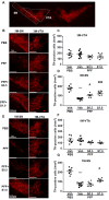
SAK3 treatment prevents the dopaminergic neuronal death in the SN but not in the VTA of PFF-injected mice. (A) The VTA and SNc region are framed by a white line. (B) The representative immunofluorescence images of TH in the VTA and SNc regions during the 3-month SAK3 treatment schedule. Scale bar: 500 μm. The number of TH-positive cells was counted in the (C) VTA and (D) SNc region (n = 6–8 per group). (E) The representative immunofluorescence images of TH in the VTA and SNc region during the 1-month SAK3 treatment schedule. Scale bar: 500 μm. The number of TH-positive cells was counted in the (F) VTA and (G) SNc region (n = 7–8 per group). Error bars represent SEM. *** p < 0.001 vs. vehicle-treated PBS-injected mice; # p < 0.05, ### p < 0.001 vs. vehicle-treated α-Syn PFF-injected mice. Abbreviations: Veh = vehicle; S = SAK3.
Figure 5
Figure 5
SAK3 administration rescues the decrease in proteasome activity in α-Syn PFF-injected mice. Proteasome activity assay using fluorogenic peptides (A,D) Suc-LLVY-AMC (chymotrypsin-like), (B,E) Bz-VGR-AMC (trypsin-like), and (C,F) Z-LLE-AMC (caspase-like) on the brain’s cortical region in both schedules (n = 6–8 per group). Error bars represent SEM. * p < 0.05, ** p < 0.01 vs. vehicle-treated PBS-injected mice; # p < 0.05, ## p < 0.01 vs. vehicle-treated α-Syn PFF-injected mice. Abbreviations: Veh = vehicle; S = SAK3.
Figure 6
Figure 6
SAK3 administration improves CaMKII-Rpt6 signaling in α-Syn PFF-injected mice. (A,D) Representative images of western blot membranes containing cortical protein, probed with antibodies against autophosphorylated CaMKII (T286), CaMKII, phosphorylated Rpt6 (S120), Rpt6, and β-actin. Quantitative analyses of (B,E) autophosphorylated CaMKII (T286) and (C,F) phosphorylated Rpt6 (S120), during the 3-month SAK3 treatment schedule (n = 4 per group) and the 1-month SAK3 treatment schedule (n = 4–6 per group). Error bars represent SEM. ** p < 0.01 vs. vehicle-treated PBS-injected mice; # p < 0.05, ## p < 0.01 vs. vehicle-treated α-Syn PFF-injected mice. Abbreviations: Veh = vehicle; S = SAK3.
Figure 7
Figure 7
Three months of SAK3 treatment improves cognitive and motor functions in α-Syn PFF-injected mice. Analyses of motor function based on the (A) rotarod task (n = 10–12 per group) and (B) beam-walking task (n = 10–12 per group) at 4, 8, and 12 weeks after mouse α-Syn PFF injection. (C) Test session of the novel object recognition task (n = 10–12 per group). (D) Test session of the step-through passive avoidance task (n = 10–12 per group). Error bars represent SEM. * p < 0.05, ** p < 0.01 vs. vehicle-treated PBS-injected mice; # p < 0.05, ## p < 0.01 vs. vehicle-treated α-Syn PFF-injected mice. Abbreviations: Veh = vehicle; S = SAK3.
Figure 8
Figure 8
High-concentration treatment with SAK3 for 1 month improves cognitive and motor functions in α-Syn PFF-injected mice. Analyses of motor function based on the (A) rotarod task (n = 10–12 per group) and (B) beam-walking task (n = 10–12 per group) at 4, 8, and 12 weeks after mouse α-Syn PFF injection. (C) Test session of the novel object recognition task (n = 10–12 per group). (D) Test session of the step-through passive avoidance task (n = 10–12 per group). Error bars represent SEM. * p < 0.05, ** p < 0.01 vs. vehicle-treated PBS injection mice; # p < 0.05, ## p < 0.01 vs. vehicle-treated α-Syn PFF-injected mice. Abbreviations: Veh = vehicle; S = SAK3.
Figure 9
Figure 9
Schematic illustration of the action mechanism of SAK3 in PD pathology. SAK3 triggers intracellular calcium influx and promotes Glu release by enhancing T-type calcium channels. Proteasome activity is increased via the CaMKII/Rpt6 signaling pathway. Facilitated proteasomal activity by SAK3 contributes to the degradation of α-Syn aggregates and potentiates synaptic plasticity. AMPAR, glutamate α-amino-4-hydroxy-5-methyl-4-isoxazolepropionic acid receptor; NMDAR, glutamate N-methyl-D-aspartate receptor; nAChR, nicotinic acetylcholine receptor.

Similar articles

Cited by

References

    1. Pillon B., Dubois B., Cusimano G., Bonnet A.M., Lhermitte F., Agid Y. Does cognitive impairment in Parkinson’s disease result from non-dopaminergic lesions? J. Neurol. Neurosurg Psychiatry. 1989;52:201–206. doi: 10.1136/jnnp.52.2.201. - DOI - PMC - PubMed
    1. Walker Z., Possin K.L., Boeve B.F., Aarsland D. Lewy body dementias. Lancet. 2015;386:1683–1697. doi: 10.1016/S0140-6736(15)00462-6. - DOI - PMC - PubMed
    1. Lippa C.F., Duda J.E., Grossman M., Hurtig H.I., Aarsland D., Boeve B.F., Brooks D.J., Dickson D.W., Dubois B., Emre M., et al. DLB and PDD boundary issues: Diagnosis, treatment, molecular pathology, and biomarkers. Neurology. 2007;68:812–819. doi: 10.1212/01.wnl.0000256715.13907.d3. - DOI - PubMed
    1. Irwin D.J., Lee V.M., Trojanowski J.Q. Parkinson’s disease dementia: Convergence of α-synuclein, tau and amyloid-β pathologies. Nat. Rev. Neurosci. 2013;14:626–636. doi: 10.1038/nrn3549. - DOI - PMC - PubMed
    1. Giehm L., Svergun D.I., Otzen D.E., Vestergaard B. Low-resolution structure of a vesicle disrupting α-synuclein oligomer that accumulates during fibrillation. Proc. Natl. Acad. Sci. USA. 2011;108:3246–3251. doi: 10.1073/pnas.1013225108. - DOI - PMC - PubMed

MeSH terms