. 2021 Jun 8;7(6):462.
doi: 10.3390/jof7060462.

Development of a Simple and Robust Whole Blood Assay with Dual Co-Stimulation to Quantify the Release of T-Cellular Signature Cytokines in Response to Aspergillus fumigatus Antigens

Affiliations

Development of a Simple and Robust Whole Blood Assay with Dual Co-Stimulation to Quantify the Release of T-Cellular Signature Cytokines in Response to Aspergillus fumigatus Antigens

Chris D Lauruschkat et al. J Fungi (Basel). .

Abstract

Deeper understanding of mold-induced cytokine signatures could promote advances in the diagnosis and treatment of invasive mycoses and mold-associated hypersensitivity syndromes. Currently, most T-cellular immunoassays in medical mycology require the isolation of mononuclear cells and have limited robustness and practicability, hampering their broader applicability in clinical practice. Therefore, we developed a simple, cost-efficient whole blood (WB) assay with dual α-CD28 and α-CD49d co-stimulation to quantify cytokine secretion in response to Aspergillus fumigatus antigens. Dual co-stimulation strongly enhanced A. fumigatus-induced release of T-cellular signature cytokines detectable by enzyme-linked immunosorbent assay (ELISA) or a multiplex cytokine assay. Furthermore, T-cell-dependent activation and cytokine response of innate immune cells was captured by the assay. The protocol consistently showed little technical variation and high robustness to pre-analytic delays of up to 8 h. Stimulation with an A. fumigatus lysate elicited at least 7-fold greater median concentrations of key T-helper cell signature cytokines, including IL-17 and the type 2 T-helper cell cytokines IL-4 and IL-5 in WB samples from patients with Aspergillus-associated lung pathologies versus patients with non-mold-related lung diseases, suggesting high discriminatory power of the assay. These results position WB-ELISA with dual co-stimulation as a simple, accurate, and robust immunoassay for translational applications, encouraging further evaluation as a platform to monitor host immunity to opportunistic pathogens.

Keywords: Aspergillus; adaptive immunity; biomarker; cytokines; immunoassay; inflammation.


Conflict of interest statement

The authors have no conflicts of interest related to this study.

Figures

Figure 1
Figure 1
Impact of co-stimulatory factors on A. fumigatus lysate-induced cytokine secretion. (a,b) WB from 10 highly mold-exposed subjects, as defined in References [3,33], was stimulated for 24 h with AfuLy in RPMI-supplemented test tubes containing α-CD28, α-CD28 plus α-CD49d, or no co-stimulatory factors (w/o co-stim.). IFN-γ (a) and IL-17 (b) concentrations were quantified in plasma supernatants by ELISA. For all results shown, the unspecific background, determined in a test tube containing the respective co-stimulation cocktail but no lysate, was deduced from antigen-induced cytokine concentrations. Corresponding test results from each donor are indicated by the same color. The Friedman test and Dunn’s multiple comparison test were used for significance testing. (c,d) WB samples from 3 additional healthy donors were stimulated as described above. Cytokine concentrations in plasma supernatants were quantified using a 21-plex Luminex assay. Individual background-adjusted concentrations of T-cellular signature cytokines (c) and selected cytokines predominantly produced by antigen-presenting cells (d) are shown. Black bars indicate means.
Figure 2
Figure 2
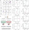
T-cell-dependent immune cell activation and cytokine release in WB-ELISA stimulation tubes. (a) Representative flow cytometry panels showing the expression of key activation markers (CD69, CD154, and/or CD107a) and IFN-γ by unstimulated, AfuLy-stimulated, and PHA-stimulated CD4+ T-helper cells, CD8+ cytotoxic T-cells, cytokine-producing CD56brightCD16 NK cells, cytotoxic CD56dimCD16+ NK cells, and CD3+CD56+ NKT cells, as determined by intracellular staining. (b) Flowchart of experimental procedures to determine the T-cell dependency of activation marker expression and cytokine response by comparison of regular and CD3-depleted WB. Ranges of CD3+ cell frequencies among leukocytes and leukocyte viability after magnetic bead-based CD3 depletion and mock depletion are provided. (c) Relative expression of the degranulation marker CD62L and activation marker CD11b on granulocytes and CD107a expression of NK cells upon AfuLy and PHA stimulation of regular (green) and CD3-depleted (red) WB, normalized to unstimulated cells. (d) Background-adjusted concentrations of IFN-γ, IL-17, IL-8, and MIP-1α (CCL3) in plasma supernatants of regular (green) and CD3-depleted (red), AfuLy- and PHA-stimulated WB. (c,d) Paired two-sided t-test; n = 4. Black bars indicate means. Abbreviations: AfuLy = A. fumigatus mycelial lysate, ex.-inh. = exocytosis inhibitors, Leuk. = leukocyte, MFI = mean fluorescence intensity, PHA = Phytohaemagglutinin, unst. = unstimulated, viab. = viability, WB = whole blood.
Figure 3
Figure 3
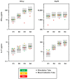
Technical and intra-individual variation of WB-ELISA. WB samples from 4 highly mold-exposed healthy subjects were stimulated with AfuLy or Aspf4 using RPMI-supplemented, α-CD28-, and α-CD49d-containing stimulation tubes as described in the Materials and Methods Section. After 24 h of stimulation, IFN-γ and IL-17 concentrations in plasma supernatants were quantified by ELISA. Individual results are indicated by black bars. For each donor and antigen (including the unspecific background control), a second set of stimulation tubes was prepared by the same operator using the same blood sample to determine technical variation (light blue diamonds). In addition, another set of stimulation tubes was injected by a different operator using blood from the same venipuncture to determine the inter-operator variation (yellow squares). Another blood draw was performed from the same subjects after a period of at least 4 weeks and cytokine concentrations (red triangle) were compared with the initial measurement (black bar) to calculate the intra-individual variation. All donors had reported no change of mold exposure profiles since the initial sampling based on our published questionnaire [33]. Colored boxes indicate the maximum range allowing for a CV of 15% (light grey) and 25% for defined protein antigens (Aspf4) or 35% for the lysate [32], respectively (dark grey). Abbreviations: AfuLy = A. fumigatus mycelial lysate, CV = coefficient of variation, D = donor, inter-oper. = inter-operator, intra-indiv. = intra-individual, Med. = median, WB = whole blood.
Figure 4
Figure 4
Robustness of WB-ELISA to pre-analytic delays. WB samples from 4 highly mold-exposed healthy donors were stimulated for 24 h with AfuLy or Aspf4 using RPMI-supplemented, α-CD28-, and α-CD49d-containing stimulation tubes, as described in the Materials and Methods Section. IFN-γ and IL-17 concentrations in plasma supernatants were quantified by ELISA. WB was injected and incubated at 37 °C either immediately (black bars) or after pre-analytic storage at room temperature for 4 (green squares), 8 (yellow squares), or 24 h (red squares). In addition, immediately injected stimulation tubes were kept at room temperature for 4 (green diamonds), 8 (yellow diamonds), or 24 h (red diamonds) prior to being transferred to 37 °C for another 24 h incubation period. Individual background-corrected cytokine concentrations for each condition are shown. Grey boxes indicate a ±2-fold change compared with the immediately incubated control sample. Abbreviations: AfuLy = A. fumigatus mycelial lysate, WB = whole blood.
Figure 5
Figure 5
Comparison of Aspergillus-induced cytokine release in patients with Aspergillus-associated lung pathologies and other chronic lung diseases. WB from 9 patients with Aspergillus-associated lung pathologies (2 CF with A. fumigatus sensitization, 3 ABPA, and 4 CPA) and a control group of 5 patients with other chronic lung diseases was stimulated with AfuLy or Aspf4 using the RPMI-supplemented, α-CD28-, and α-CD49d-containing stimulation tubes, as described in the Materials and Methods Section. Cytokine concentrations in plasma supernatants were quantified using a 21-plex Luminex assay. (a,b) Heat maps indicating median background-adjusted cytokine concentrations in the ABPA/CF/CPA patient cohort “A” and control cohort “C” depending on the antigen used for stimulation. The numeric value in the MMR column represents the median-to-median ratio between the two cohorts, with values > 1.0 indicating greater median cytokine concentrations in the ABPA/CF/CPA cohort. Δ denotes infinite median-to-median ratios (median = 0 pg/mL in the control cohort). ◊ denotes undefined median-to-median ratios (median = 0 pg/mL in both cohorts). (c,d) Heat maps summarizing individual cytokine response to AfuLy in all 14 patients. (e) Comparison of individual AfuLy-induced concentrations of selected T-helper cell signature cytokines. Medians and interquartile ranges for patients with ABPA/CF/CPA (“A”, red) and controls (“C”, grey) are indicated by black bars and colored boxes, respectively. Two-sided Mann–Whitney U test. Abbreviations: AfuLy = A. fumigatus mycelial lysate, ABPA = allergic bronchopulmonary aspergillosis, CF = cystic fibrosis, CPA = chronic pulmonary aspergillosis, Fractalk. = fractalkine, WB = whole blood.

Similar articles

Cited by

References

    1. Dewi I., van de Veerdonk F., Gresnigt M. The Multifaceted Role of T-Helper Responses in Host Defense against Aspergillus fumigatus. J. Fungi. 2017;3:55. doi: 10.3390/jof3040055. - DOI - PMC - PubMed
    1. Park S.J., Hughes M.A., Burdick M., Strieter R.M., Mehrad B. Early NK cell-derived IFN-{gamma} is essential to host defense in neutropenic invasive aspergillosis. J. Immunol. 2009;182:4306–4312. doi: 10.4049/jimmunol.0803462. - DOI - PMC - PubMed
    1. Wurster S., Weis P., Page L., Helm J., Lazariotou M., Einsele H., Ullmann A.J. Intra- and inter-individual variability of Aspergillus fumigatus reactive T-cell frequencies in healthy volunteers in dependency of mould exposure in residential and working environment. Mycoses. 2017;60:668–675. doi: 10.1111/myc.12643. - DOI - PubMed
    1. Jolink H., Hagedoorn R.S., Lagendijk E.L., Drijfhout J.W., van Dissel J.T., Falkenburg J.H., Heemskerk M.H. Induction of A. fumigatus-specific CD4-positive T cells in patients recovering from invasive aspergillosis. Haematologica. 2014;99:1255–1263. doi: 10.3324/haematol.2013.098830. - DOI - PMC - PubMed
    1. Lordan J.L., Bucchieri F., Richter A., Konstantinidis A., Holloway J.W., Thornber M., Puddicombe S.M., Buchanan D., Wilson S.J., Djukanovic R., et al. Cooperative effects of Th2 cytokines and allergen on normal and asthmatic bronchial epithelial cells. J. Immunol. 2002;169:407–414. doi: 10.4049/jimmunol.169.1.407. - DOI - PubMed

LinkOut - more resources