Review
. 2021 Jun 8;13(12):2864.
doi: 10.3390/cancers13122864.

Noninvasive Imaging Methods to Improve the Diagnosis of Oral Carcinoma and Its Precursors: State of the Art and Proposal of a Three-Step Diagnostic Process

Affiliations
Review

Noninvasive Imaging Methods to Improve the Diagnosis of Oral Carcinoma and Its Precursors: State of the Art and Proposal of a Three-Step Diagnostic Process

Antonio Romano et al. Cancers (Basel). .

Abstract

Oral squamous cell carcinoma (OSCC) is the most prevalent form of cancer of lips and oral cavity, and its diagnostic delay, caused by misdiagnosis at the early stages, is responsible for high mortality ratios. Biopsy and histopathological assessment are the gold standards for OSCC diagnosis, but they are time-consuming, invasive, and do not always enable the patient's compliance, mainly in cases of follow-up with the need for more biopsies. The use of adjunctive noninvasive imaging techniques improves the diagnostic approach, making it faster and better accepted by patients. The present review aims to focus on the most consolidated diagnostic techniques, such as vital staining and tissue autofluorescence, and to report the potential role of some of the most promising innovative techniques, such as narrow-band imaging, high-frequency ultrasounds, optical coherence tomography, and in vivo confocal microscopy. According to their contribution to OSCC diagnosis, an ideal three-step diagnostic procedure is proposed, to make the diagnostic path faster, better, and more accurate.

Keywords: Lugol’s iodine; OSCC; diagnosis; fluorescent confocal microscopy; high-frequency ultrasounds; narrow-band imaging; noninvasive diagnoses; optical coherence tomography; reflectance confocal microscopy; tissue autofluorescence; toluidine blue; virtual chromoendoscopy with magnification.


Conflict of interest statement

The authors declare no conflict of interest.

Figures

Figure 1
Figure 1
OSCC of the edentulous mucosa: (a) clinical presentation; (b) TB positivity (royal blue); (c) LI scarcely dyes the lesion, and brown-orange staining is limited to the periphery of the lesion; (d) TB and LI staining empower the identification of neoplastic areas (royal blue) and benign mucosa (brown-orange).
Figure 2
Figure 2
(a) RF at healthy mucosa: brighter at the gingiva (masticatory mucosa) than lip mucosa (covering mucosa). (b) PF, in red, due to the presence of bacterial plaque. (c,d) Reduced fluorescence due to inflammation. In these cases, anamnesis and lab tests completed the diagnosis of oral candidiasis. (e) Reduced fluorescence in a dysplasia of the cheek. (f) LF in an OSCC of the lip. (g,h) Clinical and AF pictures of an OSCC of the cheek. Inhomogeneous response to AF reveals LF at the central area of cancer (yellow dotted circle), pale reduction in fluorescence at its boundaries (white line), and PF due to necrotic tissue covered by bacterial plaque (white arrow).
Figure 3
Figure 3
Oral IPCL classification according to Takano et al. [52] (Figure designed by Maria Contaldo).
Figure 4
Figure 4
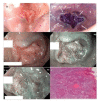
OSCC moderately differentiated. (a) Clinical photograph of a fissured exophytic lesion of the cheek. (b) Positivity to royal blue staining after TB test. Note bluish areas at the periphery of the ulceration (white arrows), expressing the persistence of suspected cells still retaining TB around the neoformation. (c) NBI imaging in white-light mode. At 60× magnification, NBI in white-light mode allows appreciating the epithelial covering of the fissured lesion. (d) At the same magnification, under narrow-band blue-green filters, NBI potentiates the capacity to appreciate subepithelial vessels. (e) At increased magnification (90×), the vascular features are more representatively seen: IPCL type IV, dilated and irregular in a brownish contest (white dotted circle), suggestive of cancer. (f) After surgery, conventional hematoxylin and eosin confirmed the diagnosis of OSCC G2, with infiltrative cancer cells and wide tumoral disarrangement of the tissue (original magnification 80×). In this case, TB and NBI help in the clinical setting before biopsy to orient the kind of surgical approach and to foresee the nature of the lesion, thus excluding traumatic injuries or non-neoplastic neoformations.
Figure 5
Figure 5
High-definition US imaging (frequency 18 MHz) of a suspicious lesion of the tongue margin. The high-definition ultrasound image shows a hypoechoic area with a thickness of 1.288 cm (TT), highlighted with the purple line, in the context of the lingual parenchyma (which does not show alterations in echogenicity). The histopathological analysis revealed the presence of OSCC of the lingual margin. Scale bar, 0.5 cm.
Figure 6
Figure 6
Leucoplakia of the cheek with dysplasia. (a) Clinical presentation of leucoplakia of the left cheek in a 41 year old male, smoker. (b) On RCM imaging, the superficial keratinization is expressed by bright areas with no identifiable cells alternated with brighter corneocyte aggregates. (c) At the upper spinous layer, polymorphic keratinocytes different in size are quite irregularly arranged and some of them show more than one nucleolus (black arrows). (d) At the lower spinous layer, the architectural pattern, slightly irregular, is intermediate between frosted glass and honeycombed, with thickened intercellular spaces. (e) Going deeper, the shadow of the connective tissue and the inflammatory exudate become visible as irregular dark areas. (f) Inflammatory cells, identified as small roundish dots, can be found in the epithelium and surrounded by dark hazy areas due to inflammatory infiltrates (white arrows). Connective tissue papillae lose their regular small roundish shape and become confluent in large elongated papillae. (g) At the submucosal layers, a strongly bright connective tissue expresses a chronic reaction, and connective bundles are irregularly arranged. Scale bars, 100 mm.
Figure 7
Figure 7
The proposed three-step diagnostic algorithm in oral pathology considering three consecutive phases: 1, data collection; 2, data integration; 3, conclusive diagnosis. (1) During the first phase, a series of noninvasive imaging techniques are performed to identify some additional diagnostic markers in addition to those obtained with conventional clinical examination and collection of anamnestic data. (2) In the second step, all data are integrated to establish the need for diagnostic biopsy. (3) In some cases, the clinical and instrumental characteristics point directly toward a conclusive diagnosis. On the other hand, when a biopsy is required, the lesion is reconsidered, with the noninvasive techniques considered, to map the lesion and suggest the correct margins and depth to the surgeons; then, the specimen is subjected to conventional histopathological evaluation and the conclusive diagnosis is obtained.

Similar articles

Cited by

References

    1. The Global Cancer Observatory. IARC. WHO Lip, Oral Cavity Cancer. [(accessed on 22 April 2021)]; Available online: https://gco.iarc.fr/today/data/factsheets/cancers/1-Lip-oral-cavity-fact....
    1. Lajolo C., Patini R., Limongelli L., Favia G., Tempesta A., Contaldo M., De Corso E., Giuliani M. Brown tumors of the oral cavity: Presentation of 4 new cases and a systematic literature review. Oral Surg. Oral Med. Oral Pathol. Oral Radiol. 2020;129:575–584.e4. doi: 10.1016/j.oooo.2020.02.002. - DOI - PubMed
    1. Favia G., Lo Muzio L., Serpico R., Maiorano E. Angiosarcoma of the head and neck with intra-oral presentation. A clinico-pathological study of four cases. Oral Oncol. 2002;38:757–762. doi: 10.1016/S1368-8375(02)00045-3. - DOI - PubMed
    1. Favia G., Lo Muzio L., Serpico R., Maiorano E. Rhabdomyoma of the head and neck: Clinicopathologic features of two cases. Head Neck. 2003;25:700–704. doi: 10.1002/hed.10266. - DOI - PubMed
    1. Lauritano D., Lucchese A., Contaldo M., Serpico R., Lo Muzio L., Biolcati F., Carinci F. Oral squamous cell carcinoma: Diagnostic markers and prognostic indicators. J. Biol. Regul. Homeost. Agents. 2016;30:169–176. - PubMed

LinkOut - more resources