. 2021 Jun 8;13(6):407.
doi: 10.3390/toxins13060407.

Colombian Scorpion Centruroides margaritatus: Purification and Characterization of a Gamma Potassium Toxin with Full-Block Activity on the hERG1 Channel

Affiliations

Colombian Scorpion Centruroides margaritatus: Purification and Characterization of a Gamma Potassium Toxin with Full-Block Activity on the hERG1 Channel

José Beltrán-Vidal et al. Toxins (Basel). .

Abstract

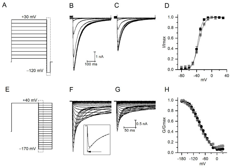
The Colombian scorpion Centruroides margaritatus produces a venom considered of low toxicity. Nevertheless, there are known cases of envenomation resulting in cardiovascular disorders, probably due to venom components that target ion channels. Among them, the humanether-à-go-go-Related gene (hERG1) potassium channels are critical for cardiac action potential repolarization and alteration in its functionality are associated with cardiac disorders. This work describes the purification and electrophysiological characterization of a Centruroides margaritatus venom component acting on hERG1 channels, the CmERG1 toxin. This novel peptide is composed of 42 amino acids with a MW of 4792.88 Da, folded by four disulfide bonds and it is classified as member number 10 of the γ-KTx1 toxin family. CmERG1 inhibits hERG1 currents with an IC50 of 3.4 ± 0.2 nM. Despite its 90.5% identity with toxin ɣ-KTx1.1, isolated from Centruroides noxius, CmERG1 completely blocks hERG1 current, suggesting a more stable plug of the hERG channel, compared to that formed by other ɣ-KTx.

Keywords: Centruroides margaritatus; CmERG1; CnERG1; ERG channel; ERG toxin; Electrophysiology.


Conflict of interest statement

The authors declare no conflict of interest.

Figures

Figure 1
Figure 1
Purification of potassium channel toxins from the venom of Centruroides margaritatus. (A) Venom separation by RP-FPLC resulting in 41 fractions. Fraction F23 is active on hERG1 channels and is indicated by the red arrow. The inset shows an example of the hERG1 current (black line) and the blocking effect of F23 (red line). (B) Re-purification of the fraction F23; the pure peptide active on hERG1 is indicated by the asterisk and corresponds to the one with MW 4792.88 Da and called CmERG1. (CE) Isolation of CmERG1 by means of a three-step protocol. First, gel filtration leads to three principal fractions FI, FII and FIII (C). From these, fraction FII was further separated by cation-exchange chromatography into 10 sub-fractions (D). Toxin CmERG1 was isolated by RP-HPLC from fractions FII.6 with a retention time of 27.4 min (indicated with a star in E). In C and D, the red arrow indicates the fraction separated in the subsequent step.
Figure 2
Figure 2
Alignment of CmERG1 with similar ERG toxins from the family γKTx1. Cysteines are highlighted in yellow. The important residues for the CnERG1-hERG1 channel interaction, according to [19,20], are indicated in magenta. Points indicate identical positions to those in CmERG1.
Figure 3
Figure 3
CmERG1 toxin blocking features. (A) Dose-response curve. Normalized peak currents were plotted versus the logarithm of toxin concentration. The grey line represents the best fit with a logistic equation giving an IC50 and a slope of 3.4 ± 0.2 nM and 1.1 ± 0.05, respectively. (B) A representative hERG1 current in control (black line) and after application of CmERG1 toxin at 300 nM (light grey trace) and 3 nM (grey trace and red trace for resized current). (C) Time course of block during toxin application at 1 µM and during washout. Stimulus was a single depolarized step at 60 mv followed by a hyperpolarized step at −120 mV, applied every 5 s. (n = 3 for data recorded at 1 μM and 10 nM; n = 5 for data at 300 nM; n = 4 for data at 100 and 30 nM; n = 6 for data at 1 and 3 nM).
Figure 4
Figure 4
Effect of CmERG1 at 4 nM on the voltage dependence of activation and inactivation. (A) Stimulation protocol used to study the voltage dependence of activation. Tail currents were elicited by 5 s preconditioning steps ranging from +30 to −80 mV, followed by a 500 ms pulse at −120 mV. (B,C) typical example of tail currents recorded in control conditions and during application of CmERG1, respectively. (D) Voltage dependence of activation. Normalized peak currents are plotted against the voltage of the preconditioning pulses in control conditions (black squares) and in the presence of 4 nM CmERG1 (grey squares). Black lines are the best fit obtained to a Boltzmann equation giving V1/2 for activation of −37.5 ± 1.4 mV and −34.8 ± 0.8 mV and slope of 6.0 ± 0.3 and 8 ± 0.7 for the control and in the presence of 4 nM CmERG1, respectively (mean ± SE and n = 5, analyzed by paired sample t-test at 0.05 level). (E) Stimulation used to study the voltage dependence of inactivation. Currents were elicited by a 1 s preconditioning pulse at 40 mV followed by depolarization steps ranging from −170 mV to 40 mV. Currents recorded at potentials lower than −90 mV were corrected for deactivation as shown in the inset of panel (F). The deactivation process was fitted by a single exponential (grey dash line) extrapolated to the zero point of the depolarization step, indicated by the arrow. (F,G) typical example of currents recorded in control conditions and in presence of 4 nM CmERG1 respectively. (H) Voltage dependence of inactivation. Conductance was extrapolated from corrected peak currents, normalized, and then plotted versus voltage. Black lines are the best fit obtained by a Boltzmann equation giving V1/2 for inactivation of −77.6 ± 3.6 mV and −79 ± 1.3 mV and slope of 28.2 ± 2.2 and 29 ± 1.5 for control conditions and in presence of 4 nM CmERG1, respectively (mean ± SE and n = 5, analyzed by paired sample t-test at 0.05 level).
Figure 5
Figure 5
(A,B): Two possible binding modes that block the access to the selectivity filter. The channel pore is shown as an orange ribbon and the toxins as green ribbons. The contact volume or “seal” between the channel and the toxin (with a 4.5 Å cutoff) is shown as a translucent purple surface. Residues in the toxins that have been shown to affect binding or that are different between CnERG1 and CmERG1 (see Figure 2) are shown as spheres in CPK colors (C in cyan, O in red, N in blue, S in yellow). (A) In a high affinity pose, CmERG1 K13 penetrates the entry to the selectivity filter (marked by the orange spheres labeled as G628), flanked by Y14, F37, and F36 (behind K13 and F37), making a hydrophobic and cationic plug. (B) In a lower affinity pose for CnERG1, Y14 lies directly over the entry to the selectivity filter (marked by the orange spheres labeled as G628), while K13 engages in hydrogen bonds with two of the G628 carbonyls. (C) Top view of the CnERG1 complex, showing that one toxin is enough to fill the pore entrance of the channel.
Figure 6
Figure 6
Channel-CmERG1 interactions in a high affinity pose for residues that impair binding when mutated (from Figure 2). The channel is displayed as an orange ribbon and the residues in licorice and CPK colors. Toxin residues include hydrogen atoms; channel residues are labeled in boldface and italics. Residues that interact with the selectivity filter; (A) K13 engages in hydrogen bonds with three of the four mainchain carbonyls of the F627 residues and van der Waals interactions with G626 and G628, blocking the pore. (B) Y14 is nested in a crevice on the side of the entrance to the selectivity filter, making van der Waals contacts with residues from two adjacent subunits. (C) F37 stacks in a T conformation against W585 and makes van der Waals contacts with the carbonyl of G628 at the entrance of the selectivity filter. Residues that interact with turret residues and contribute to the “seal” between the toxin and the channel: (D) F17 interacts with Y597 in a T conformation. (E) M35 is surrounded by the sidechains of S581, R582, and N588. (F) Q18 stacks against the peptide bond between Y597 and P596, and hydrogen bonds to R582 from the adjacent subunit. This same R also hydrogen bonds to the carbonyl of P596.
Figure 6
Figure 6
Channel-CmERG1 interactions in a high affinity pose for residues that impair binding when mutated (from Figure 2). The channel is displayed as an orange ribbon and the residues in licorice and CPK colors. Toxin residues include hydrogen atoms; channel residues are labeled in boldface and italics. Residues that interact with the selectivity filter; (A) K13 engages in hydrogen bonds with three of the four mainchain carbonyls of the F627 residues and van der Waals interactions with G626 and G628, blocking the pore. (B) Y14 is nested in a crevice on the side of the entrance to the selectivity filter, making van der Waals contacts with residues from two adjacent subunits. (C) F37 stacks in a T conformation against W585 and makes van der Waals contacts with the carbonyl of G628 at the entrance of the selectivity filter. Residues that interact with turret residues and contribute to the “seal” between the toxin and the channel: (D) F17 interacts with Y597 in a T conformation. (E) M35 is surrounded by the sidechains of S581, R582, and N588. (F) Q18 stacks against the peptide bond between Y597 and P596, and hydrogen bonds to R582 from the adjacent subunit. This same R also hydrogen bonds to the carbonyl of P596.
Figure 7
Figure 7
Channel-CnERG1 interactions in a high affinity pose for residues that impair binding when mutated (from Figure 2). The channel is displayed as an orange ribbon and the residues in licorice and CPK colors. Toxin residues include hydrogen atoms; channel residues are labeled in boldface and italics. Residues that interact with the selectivity filter: (A) K13 engages in hydrogen bonds with three of the four mainchain carbonyls of the F627 residues and van der Waals interactions with G626 and G628, blocking the pore. (B) Y14 is nested in a crevice on the side of the entrance to the selectivity filter, making van der Waals contacts with residues from two adjacent subunits and a hydrogen bond to W585 or S631. (C) F37 stacks in a parallel conformation against W585 and N588, while making a hydrogen bond with Y597 with its carbonyl. Residues that interact with turret residues and contribute to the “seal” between the toxin and the channel: (D) Y17 interacts with Y597 in a T conformation. (E): Q18 stacks against Y597 and the backbone of K595, and hydrogen bonds to H578 and R582 from the adjacent subunit. (F) Q21 is nested against the hydrogen bond between R582 and N598, engaging also in a T interaction with H578. (G) M35 is surrounded by the sidechains of S581, R582, Y597, and N588.
Figure 7
Figure 7
Channel-CnERG1 interactions in a high affinity pose for residues that impair binding when mutated (from Figure 2). The channel is displayed as an orange ribbon and the residues in licorice and CPK colors. Toxin residues include hydrogen atoms; channel residues are labeled in boldface and italics. Residues that interact with the selectivity filter: (A) K13 engages in hydrogen bonds with three of the four mainchain carbonyls of the F627 residues and van der Waals interactions with G626 and G628, blocking the pore. (B) Y14 is nested in a crevice on the side of the entrance to the selectivity filter, making van der Waals contacts with residues from two adjacent subunits and a hydrogen bond to W585 or S631. (C) F37 stacks in a parallel conformation against W585 and N588, while making a hydrogen bond with Y597 with its carbonyl. Residues that interact with turret residues and contribute to the “seal” between the toxin and the channel: (D) Y17 interacts with Y597 in a T conformation. (E): Q18 stacks against Y597 and the backbone of K595, and hydrogen bonds to H578 and R582 from the adjacent subunit. (F) Q21 is nested against the hydrogen bond between R582 and N598, engaging also in a T interaction with H578. (G) M35 is surrounded by the sidechains of S581, R582, Y597, and N588.

Similar articles

Cited by

References

    1. Ward M.J., Ellsworth S.A., Nystrom G.S. A global accounting of medically significant scorpions: Epidemiology, major toxins, and comparative resources in harmless counterparts. Toxicon. 2018;151:137–155. doi: 10.1016/j.toxicon.2018.07.007. - DOI - PubMed
    1. Rokyta D.R., Ward M.J. Venom-gland transcriptomics and venom proteomics of the black-back scorpion (Hadrurus spadix) reveal detectability challenges and an unexplored realm of animal toxin diversity. Toxicon. 2017;128:23–37. doi: 10.1016/j.toxicon.2017.01.014. - DOI - PubMed
    1. De Armas L.F., Sarmiento D.L., Flórez E.L. Composición del género Centruroides marx, 1890 (Scorpiones buthidae) en Colombia, con la descripción de una nueva especie. Boletín Soc. Entomológica Aragonsa. 2012;50:105–114.
    1. Guerrero-Vargas J.A., Rodríguez Buitrago J.R., Ayerbe S., Flórez Daza E., Beltrán Vidal J.T. Scorpionism and Dangerous Species of ColombiaColombia. In: Gopalakrishnakone P., Possani L.D., F. Schwartz E., Rodríguez de la Vega R.C., editors. Scorpion Venoms. Springer; Dordrecht, The Netherlands: 2015. pp. 245–272. - DOI
    1. Marinkelle C.J., Stahnke H.L. Toxicological and clinical studies on Centruroides margaritatus (Gervais), a common scorpion in western Colombia. J. Med. Entomol. 1965;2:197–199. doi: 10.1093/jmedent/2.2.197. - DOI - PubMed

Publication types

MeSH terms

LinkOut - more resources