. 2021 Jun 8;22(12):6200.
doi: 10.3390/ijms22126200.

Genetic Dissection of Vps13 Regulation in Yeast Using Disease Mutations from Human Orthologs

Affiliations

Genetic Dissection of Vps13 Regulation in Yeast Using Disease Mutations from Human Orthologs

Jae-Sook Park et al. Int J Mol Sci. .

Abstract

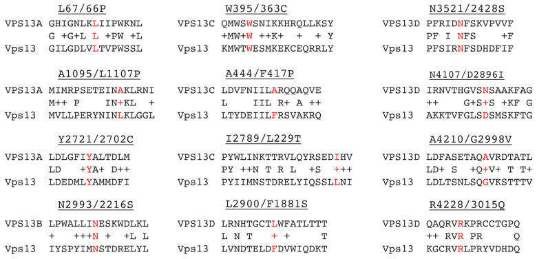
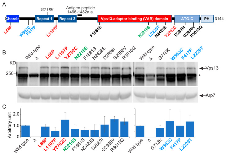
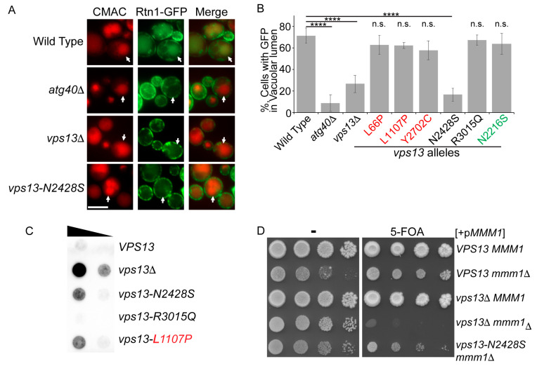
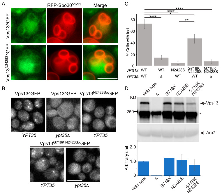
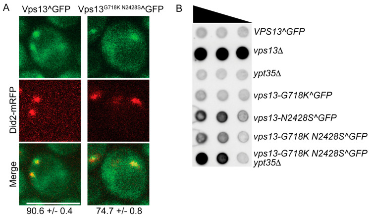
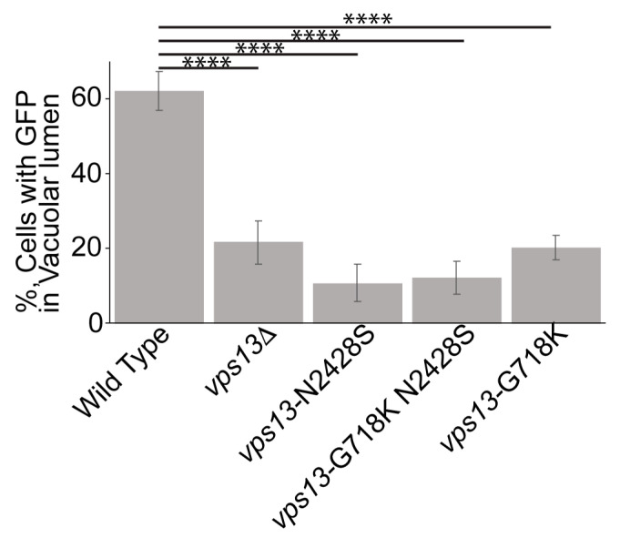
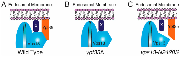
The VPS13 family of proteins have emerged as key players in intracellular lipid transport and human health. Humans have four different VPS13 orthologs, the dysfunction of which leads to different diseases. Yeast has a single VPS13 gene, which encodes a protein that localizes to multiple different membrane contact sites. The yeast vps13Δ mutant is pleiotropic, exhibiting defects in sporulation, protein trafficking, endoplasmic reticulum (ER)-phagy and mitochondrial function. Non-null alleles resulting from missense mutations can be useful reagents for understanding the multiple functions of a gene. The exceptionally large size of Vps13 makes the identification of key residues challenging. As a means to identify critical residues in yeast Vps13, amino acid substitution mutations from VPS13A, B, C and D, associated with human disease, were introduced at the cognate positions of yeast VPS13, some of which created separation-of-function alleles. Phenotypic analyses of these mutants have revealed that the promotion of ER-phagy is a fourth, genetically separable role of VPS13 and provide evidence that co-adaptors at the endosome mediate the activity of VPS13 in vacuolar sorting.

Keywords: ER-phagy; Vps13 adaptor; chorea-acanthocytosis; protein trafficking.


Conflict of interest statement

The authors declare no conflict of interest.

Figures

Figure 1
Figure 1
Alignments of regions of different human VPS13 paralogs and yeast Vps13. Labels have the format “Mammalian amino acid and position/yeast cognate amino acid and position, ending with the amino acid in the mammalian gene mutant”. The cognate amino acids are highlighted in red. All the alignments were produced by BLAST search except for the regions around VPS13B N2993 and VPS13D L2900, which were based on the VAB repeat sequences defined in [21].
Figure 2
Figure 2
Steady-state levels of yeast Vps13 proteins carrying cognate mutations of various human VPS13 paralogs (A) The position of VPS13 cognate mutations with respect to domains of the Vps13 protein. Domains are not drawn to scale. Mutations from the different human VPS13 paralogs are color coded: VPS13A, red; VPS13B, green; VPS13C, blue; VPS13D, black. G718K is a suppressor mutation identified in yeast [13]. The black bar indicates the location of the peptide used to generate the Vps13 antibody (a.a. = amino acid). (B) Immunoblot analysis. Protein extracts from the following strains: wild type (BY4741), vps13Δ (KO1), vps13-L66P (RP201), vps13-L1107P (RP202), vps13-Y2702C (RP203), vps13-N2216S (JSP770), vps13-F1881S (JSP786), vps13-N2428S (JSP757), vps13-D2869I (JSP787), vps13-G2998V (JSP788), vps13-R3015Q (JSP758), vps13-G718K (JSP549), vps13-W363C (RP206), vps13-F417P (RP207), vps13-L2229T (JSP729) were probed with anti-Vps13 and anti-Arp7 antibodies. The white line indicates two different blots. The asterisk indicates a cross-reacting band. Full blot is shown in Supplementary Figure S1. (C) Quantification of Vps13 mutant proteins relative to wild-type Vps13 ([Vps13-X/Arp7]/[Vps13/Arp7]). Graphs show the average of three independent experiments. Error bars indicate one standard deviation.
Figure 3
Figure 3
Characterization of the vps13-N2428S allele. (A) ER-phagy: cells at mid-log phase were treated with rapamycin for 16 h and stained with the dye CMAC to indicate the position of the vacuole. ER-phagy results in the co-localization of the cortical ER protein, Rtn1-GFP, with the vacuole. Wild type (GCY5), atg40Δ (JSP845), vps13Δ (JSP846) and vps13-N2428S (JSP851). Arrows highlight the positions of the vacuole in representative cells as shown by CMAC fluorescence (in red). Scale bar = 5 µm. (B) The fraction of cells exhibiting Rtn1-GFP fluorescence in the vacuolar lumen after rapamycin treatment in different VPS13 mutants; strains as in (A), plus vps13-L66P (JSP847), vps13-L1107P (JSP848), vps13-Y2702C (JSP849), vps13-R3015Q (JSP852), and vps13-N2216S (JSP854). Numbers are the averages of at least three independent experiments with over 150 cells per strain scored in each experiment. Error bars indicate one standard deviation. **** indicates p < 0.0001, Student’s t-test, n.s. not significant. (C) CPY secretion assay. Cells were grown to exponential phase in liquid YPD, adjusted to equalize cell numbers between cultures and ten-fold dilutions were spotted onto SD complete medium. A nitrocellulose membrane was placed over the cells and the plates were incubated overnight at 30 °C. The filter was then rinsed and probed with anti-CPY antibodies. Strains used were wild type (BY4741), vps13Δ (KO1), vps13-N2428S (JSP757), vps13-R3015Q (JSP758), and vps13-L1107P (RP202). (D) Synthetic lethality assay with mmm1Δ. Strains of the indicated genotype carrying pRS316-MMM1 were grown overnight at 30 °C in YPD medium; wild type (BY4741), mmm1Δ (JSP441), vps13Δ (KO1), vps13Δ mmm1Δ (JSP443) and vps13-N2428S mmm1Δ (JSP759). Tenfold serial dilutions were spotted onto SD complete medium with or without 5-FOA and grown at 30 °C for 4 days. Color code for allele designations as in Figure 2.
Figure 4
Figure 4
Localization of Vps13N2428S^GFP under various conditions. (A) Sporulation: VPS13^GFP/vps13Δ (JSYD4) and vps13-N2428S^GFP/vps13Δ (JSYD12) diploids carrying the prospore membrane marker RFP-Spo2051-91 (pRS426-R20) were transferred to SPO medium and proteins were visualized by fluorescence microscopy [38]. Representative cells are shown from over 100 cells with prospore membranes. (B) VPS13^GFP (JSP497), VPS13^GFP ypt35Δ (JSP861), vps13-N2428S^GFP (JSP816), vps13-G718K N2428S^GFP (JSP832), or vps13-G718K N2428S^GFP ypt35Δ (JSP856) were grown to log-phase in 2% glucose and then analyzed for GFP fluorescence. Scale bars = 5 µm. (C) Quantification of GFP foci in vegetative cells in glucose. Percentages are the average of at least three independent experiments with over 130 cells per strain scored in each experiment. The error bars indicate one standard deviation. **** indicates difference is significant to p < 0.0001, ** p < 0.01, * p < 0.05 by Student’s t-test. WT = wild type. (D) Western blot showing levels of the wild type and mutant GFP fusion proteins. Lower panel, levels of the mutant proteins relative to wild type, average of three experiments. Full blot is shown in Supplementary Figure S2.
Figure 5
Figure 5
The intragenic vps13-G718K mutation restores endosomal localization of Vps13G718K N2428S and promotes CPY sorting. (A) Colocalization of Vps13^GFP proteins with endosomes. Strains carrying VPS13^GFP (JSP512) or vps13-G718K N2428S^GFP (JSP864) containing the endosomal marker, DID2-mRFP, were grown in SD complete medium and collected at mid-log phase for microscopy [39]. Scale bars = 5 µm. Numbers at the bottom indicate the percentage of Vps13 foci that co-localized with the Did2-mRFP marker (±one standard deviation). Two experiments with at least 100 foci were scored for each experiment). (B) CPY sorting. The indicated strains were assayed for CPY sorting as described in Figure 4 except that 4-fold serial dilutions were used; VPS13^GFP (JSP497), vps13Δ (KO1), ypt35Δ (KO6), vps13-G718K^GFP (JSP531), vps13-N2428S^GFP (JSP816), vps13-G718K N2428S^GFP (JSP832) and vps13-G718K N2428S^GFP ypt35Δ (JSP856).
Figure 6
Figure 6
The vps13-G718K mutant is defective in ER-phagy. Quantification of ER-phagy in different strains: wild type (GCY5), vps13Δ (JSP846), vps13-N2428S (JSP851), vps13-G718K N2428S (JSP881), and vps13-G718K (JSP883). Percentages shown are the averages of at least four independent experiments and over 150 cells per strain were scored in each experiment. Error bars are one standard deviation. **** indicates comparison to wild type, p < 0.0001.
Figure 7
Figure 7
Two-adaptor model for Vps13 binding to the endosome. (A) In wild-type cells, Vps13 is stably recruited to the endosome by a strong interaction with Ypt35 and a weak interaction with a second, hypothetical protein. Both proteins interact with the Vps13 VAB domain. Interaction with X is required for proper CPY sorting. (B) In ypt35Δ cells, only the weak interaction is present. This is sufficient for CPY sorting but not for a stable localization to the endosome. (C) In vps13-2428S cells, both interactions are disrupted and both localization and CPY sorting are lost.

Similar articles

Cited by

References

    1. Dziurdzik S.K., Conibear E. The Vps13 Family of Lipid Transporters and Its Role at Membrane Contact Sites. Int. J. Mol. Sci. 2021;22:2905. doi: 10.3390/ijms22062905. - DOI - PMC - PubMed
    1. Gauthier J., Meijer I.A., Lessel D., Mencacci N.E., Krainc D., Hempel M., Tsiakas K., Prokisch H., Rossignol E., Helm M.H., et al. Recessive mutations in VPS13D cause childhood onset movement disorders. Ann. Neurol. 2018;83:1089–1095. doi: 10.1002/ana.25204. - DOI - PubMed
    1. Kolehmainen J., Black G.C., Saarinen A., Chandler K., Clayton-Smith J., Traskelin A.L., Perveen R., Kivitie-Kallio S., Norio R., Warburg M., et al. Cohen syndrome is caused by mutations in a novel gene, COH1, encoding a transmembrane protein with a presumed role in vesicle-mediated sorting and intracellular protein transport. Am. J. Hum. Genet. 2003;72:1359–1369. doi: 10.1086/375454. - DOI - PMC - PubMed
    1. Lesage S., Drouet V., Majounie E., Deramecourt V., Jacoupy M., Nicolas A., Cormier-Dequaire F., Hassoun S.M., Pujol C., Ciura S., et al. Loss of VPS13C Function in Autosomal-Recessive Parkinsonism Causes Mitochondrial Dysfunction and Increases PINK1/Parkin-Dependent Mitophagy. Am. J. Hum. Genet. 2016;98:500–513. doi: 10.1016/j.ajhg.2016.01.014. - DOI - PMC - PubMed
    1. Rampoldi L., Dobson-Stone C., Rubio J.P., Danek A., Chalmers R.M., Wood N.W., Verellen C., Ferrer X., Malandrini A., Fabrizi G.M., et al. A conserved sorting-associated protein is mutant in chorea-acanthocytosis. Nat. Genet. 2001;28:119–120. doi: 10.1038/88821. - DOI - PubMed

MeSH terms

Substances

LinkOut - more resources