. 2021 Jun 8;8(6):79.
doi: 10.3390/bioengineering8060079.

3D Printing of Thermoresponsive Hydrogel Laden with an Antimicrobial Agent towards Wound Healing Applications

Affiliations

3D Printing of Thermoresponsive Hydrogel Laden with an Antimicrobial Agent towards Wound Healing Applications

Martyna Nizioł et al. Bioengineering (Basel). .

Abstract

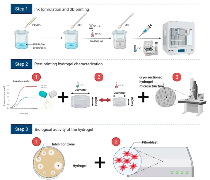
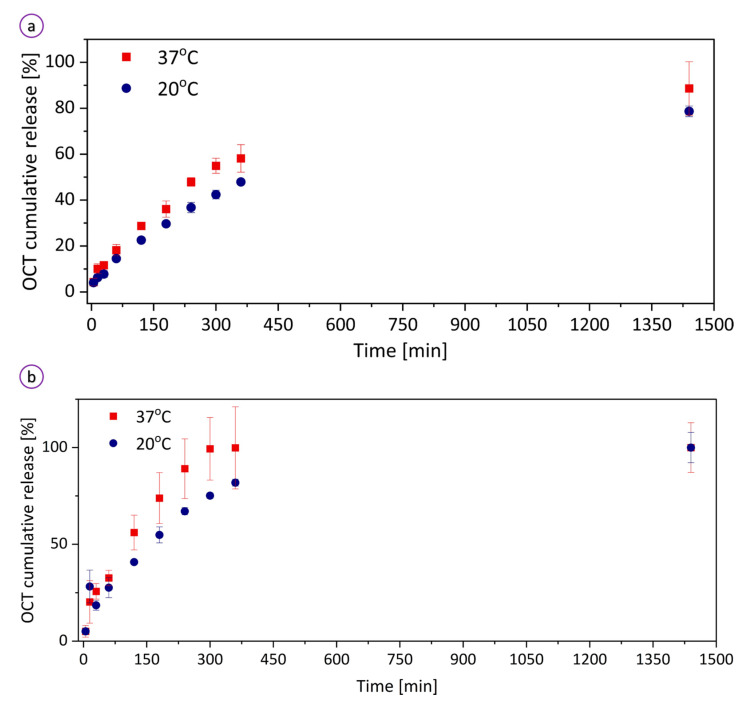
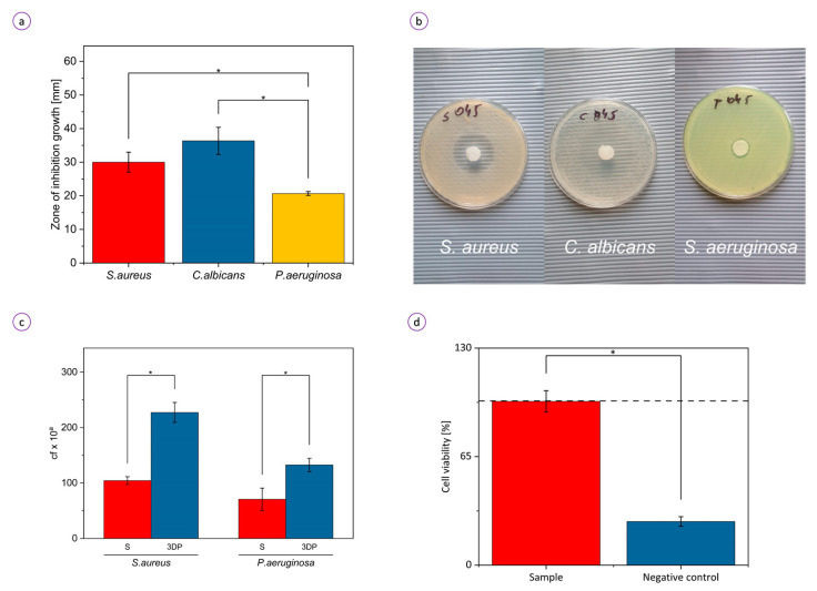
Thermoresponsive hydrogel-based wound dressings with an incorporated antimicrobial agent can be fabricated employing 3D printing technology. A novel printable ink containing poly(N-isopropylacrylamide) (PNIPAAm) precursors, sodium alginate (ALG), methylcellulose (MC) that is laden with a mixture of octenidine dihydrochloride and 2-phenoxyethanol (Octenisept®, OCT) possess accurate printability and shape fidelity. This study also provides the protocol of ink's use for the 3D printing of hydrogel scaffolds. The hydrogel's physicochemical properties and drug release profiles from the hydrogel specimens to the external solution have been determined at two temperatures (20 and 37 °C). The release test showed a sustained OCT delivery into ultrapure water and the PBS solution. The temperature-responsive hydrogel exhibited antimicrobial activity against Staphylococcus aureus, Candida albicans, and Pseudomonas aeruginosa and demonstrated non-cytotoxicity towards fibroblasts. The thermoresponsive behavior along with biocompatibility, antimicrobial activity, and controlled drug release make this hydrogel a promising class of materials for wound dressing applications.

Keywords: additive manufacturing; biocompatible; printability; stimuli-responsive; wound patch.


Conflict of interest statement

The authors declare no conflict of interest.

Figures

Figure 1
Figure 1
Summary of the procedure: Step 1 demonstrates all steps of the ink formulation and 3D printing including the addition of hydrogel precursors and pre-printing stages such as bubble removal and pouring the ink into a printing syringe; Step 2 presents the main characterization stages that include (1) OCT release profile determination, (2) swelling tests, and (3) SEM observations of the hydrogel cross-sectioned structure; in Step 3, the hydrogel is characterized for its antimicrobial activity and biocompatibility. Created using Biorender.com.
Figure 2
Figure 2
Rheological properties of the hydrogel ink: (a) Frequency and (b) strain (at a frequency of 1 Hz) dependence of storage and loss moduli. Inset in (a) shows the magnified plot for the frequency of 0–10 Hz; (c) Complex viscosity of the hydrogel precursor. Inset shows the magnified plot for the shear rate of 0–5 1/s; (d) shear stress vs. shear rate plot.
Figure 3
Figure 3
Hydrogel characterization: (a) ATR-FTIR spectrum of the 3D-printed sample with (top) and without (bottom) OCT; (b,c) cryo-SEM images of the cross-sectional microstructure of the specimens. Bars correspond to 40 and 5 μm, respectively; (d) stress–strain curve of the hydrogel; (e) Young’s modulus and compression strength of the material.
Figure 4
Figure 4
Three-dimensional (3D) printability and temperature-induced behavior of the hydrogel: (a) 3D-printed thermoresponsive tube demonstrates high printability of the ink. The combination of 3D printing and ink allows for the fabrication of hollow tubular structures, whose diameter alters in response to temperature change; (b) 3D-printed flower-like object with thermoresponsive petals and non-active core; (c) 3D-printed thermoresponsive hydrogel propeller; (4) 3D-printed thermoresponsive hydrogel disc. Figure (bd) demonstrate that objects printed from the ink can be programmed to exhibit different actuation behaviors at different temperatures (42 and 10 °C). Bars correspond to 1 cm. The hydrogel swelling rates at temperatures 20 and 37 °C in water (e) and PBS (f); (g) Temperature-activated swelling (20 °C) and de-swelling (37 °C) cycles of the sample measured as a change of specimen height, diameter, and mass. The values are normalized to those at a temperature of 37 °C.
Figure 5
Figure 5
OCT release profiles from the hydrogel to (a) water and (b) PBS (pH of 7.4) at temperatures of 20 and 37 °C.
Figure 6
Figure 6
Biological activity of the hydrogel: (a) average diameter of inhibition zones of S. aureus, C. albicans, and P. aeruginosa and (b) corresponding photos of the dishes; (c) ability of S. aureus and P. aeruginosa to colonize the molded (S) and 3D-printed (3DP) hydrogel samples. An exponent “a” corresponds to 9 and 6 for S. aureus and P. aeruginosa, respectively; “cf” means colony-forming; (d) viability of fibroblasts exposed to medium conditioned with extracts from the analyzed sample and ethanol (negative control). Absorbance at 490 nm measured for unexposed fibroblasts (C) was considered 100% (black dashed line); * p < 0.05.

References

    1. Qasim M., Haq F., Kang M.H., Kim J.H. 3D printing approaches for cardiac tissue engineering and role of immune modulation in tissue regeneration. Int. J. Nanomed. 2019;14:1311–1333. doi: 10.2147/IJN.S189587. - DOI - PMC - PubMed
    1. Blacklow S.O., Li J., Freedman B.R., Zeidi M., Chen C., Mooney D.J. Bioinspired mechanically active adhesive dressings to accelerate wound closure. Sci. Adv. 2019;5:eaaw3963. doi: 10.1126/sciadv.aaw3963. - DOI - PMC - PubMed
    1. Yang M., He S., Su Z., Yang Z., Liang X., Wu Y. Thermosensitive Injectable Chitosan/Collagen/β-Glycerophosphate Composite Hydrogels for Enhancing Wound Healing by Encapsulating Mesenchymal Stem Cell Spheroids. ACS Omega. 2020;5:21015–21023. doi: 10.1021/acsomega.0c02580. - DOI - PMC - PubMed
    1. Wang X., Jiang B., Sun H., Zheng D., Zhang Z., Yan L., Li E., Wu Y., Xu R.-H. Noninvasive application of mesenchymal stem cell spheres derived from hESC accelerates wound healing in a CXCL12-CXCR4 axis-dependent manner. Theranostics. 2019;9:6112–6128. doi: 10.7150/thno.32982. - DOI - PMC - PubMed
    1. Qian Z., Wang H., Bai Y., Wang Y., Tao L., Wei Y., Fan Y., Guo X., Liu H. Improving Chronic Diabetic Wound Healing through an Injectable and Self-Healing Hydrogel with Platelet-Rich Plasma Release. ACS Appl. Mater. Interfaces. 2020;12:55659–55674. doi: 10.1021/acsami.0c17142. - DOI - PubMed

LinkOut - more resources