. 2021 Jun 23;22(13):6712.
doi: 10.3390/ijms22136712.

CD163 as a Potential Biomarker of Monocyte Activation in Ischemic Stroke Patients

Affiliations

CD163 as a Potential Biomarker of Monocyte Activation in Ischemic Stroke Patients

Rosaria Greco et al. Int J Mol Sci. .

Abstract

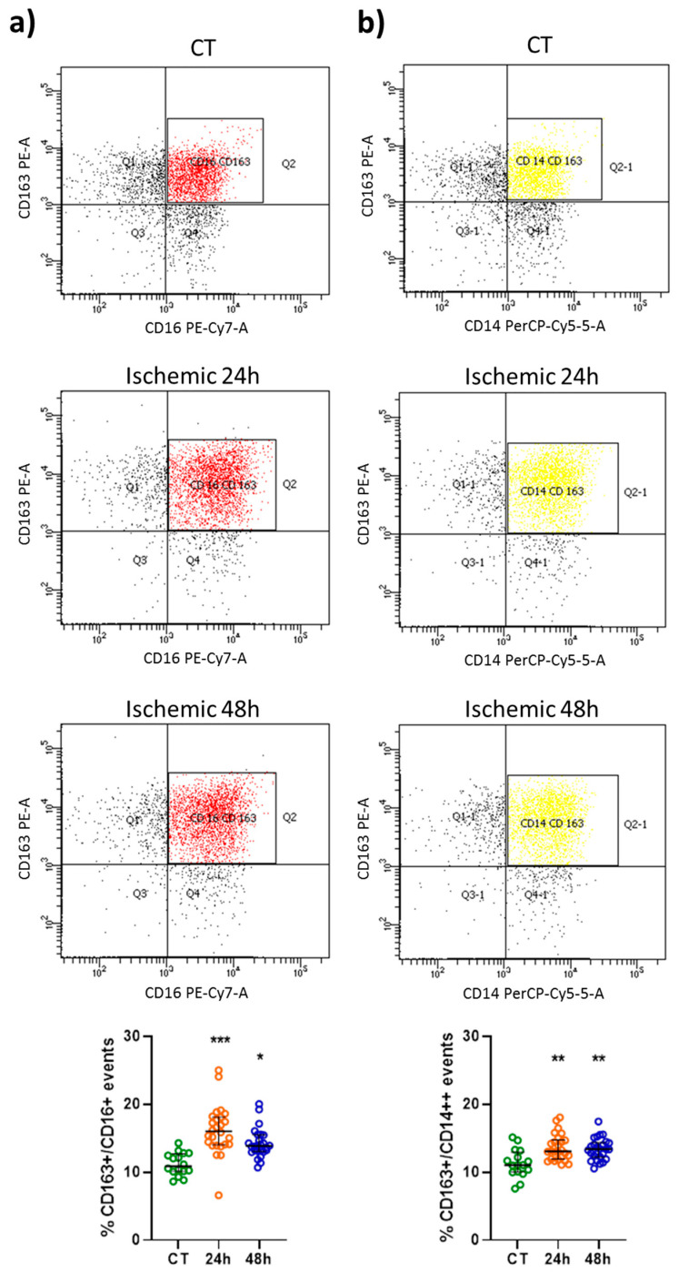
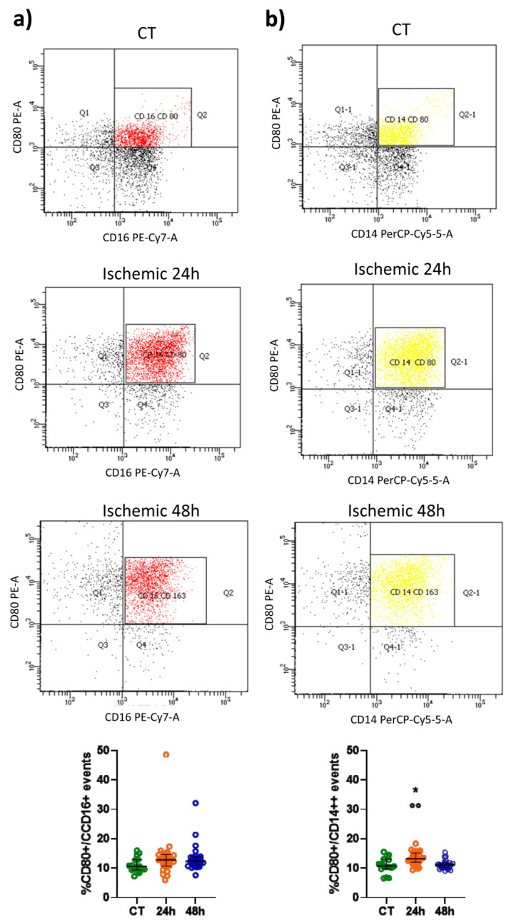
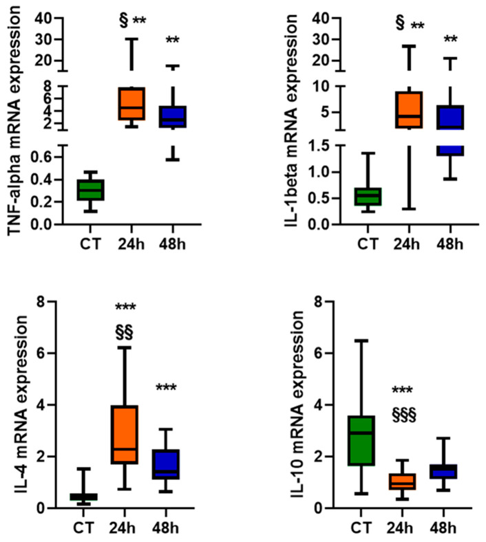
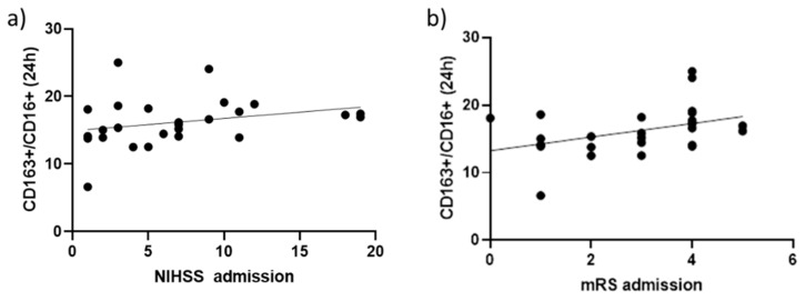
In ischemic stroke patients, a higher monocyte count is associated with disease severity and worse prognosis. The complex correlation between subset phenotypes and functions underscores the importance of clarifying the role of monocyte subpopulations. We examined the subtype-specific distribution of the CD163+ and CD80+ circulating monocytes and evaluated their association with the inflammatory status in 26 ischemic stroke patients and 16 healthy controls. An increased percentage of CD163+/CD16+ and CD163+/CD14++ events occurred 24 and 48 h after a stroke compared to the controls. CD163+ expression was more pronounced in CD16+ non-classical and intermediate monocytes, as compared to CD14+ classical subtype, 24 h after stroke. Conversely, the percentage of CD80+/CD16+ events was unaffected in patients; meanwhile, the percentage of CD80+/CD14+ events significantly increased only 24 h after stroke. Interleukin (IL)-1beta, TNF-alpha, and IL-4 mRNA levels were higher, while IL-10 mRNA levels were reduced in total monocytes from patients versus controls, at either 24 h or 48 h after stroke. The percentage of CD163+/CD16+ events 24 h after stroke was positively associated with NIHSS score and mRS at admission, suggesting that stroke severity and disability are relevant triggers for CD163+ expression in circulating CD16+ monocytes.

Keywords: CD163+; CD80+; acute ischemic stroke; cytokines; peripheral blood monocytes.


Conflict of interest statement

The authors declare no conflict of interest.

Figures

Figure 1
Figure 1
Representative flow cytometry density plots and scatter plots showing the percentage of CD163+/CD16+ (red) (a) and CD163+/CD14++ (yellow) (b) events in healthy subjects (CT; n = 16) and in patients 24 and 48 h after an ischemic stroke (n = 26). Data are shown as the median and interquartile range. A Kruskal–Wallis test, followed by Dunn’s post-hoc test: * p < 0.05, ** p < 0.01, and *** p < 0.001 vs. CT.
Figure 2
Figure 2
Representative flow cytometry density plots and scatter plots showing the percentage of CD80+/CD16+ (red) (a) and CD80+/CD14++ (yellow) (b) events in healthy subjects (CT; n = 16) and in patients 24 and 48 h after an ischemic stroke (n = 26). Data are shown as the median and interquartile range. A Kruskal–Wallis test, followed by Dunn’s post-hoc test: * p < 0.05 vs. CT; Wilcoxon’s signed-rank test: °° p < 0.01 vs. 48 h.
Figure 3
Figure 3
Cytokines’ mRNA levels, expressed as relative quantification (RQ) in the monocytes of healthy subjects (CT; n = 16) and patients 24 and 48 h after an ischemic stroke (n = 24–26). Data are shown as the median and the minimum and maximum values. A Kruskal–Wallis test, followed by Dunn’s post-hoc test: ** p < 0.01 and *** p < 0.001 vs. CT; Wilcoxon’s signed-rank test: § p < 0.05, §§ p < 0.01, and §§§ p < 0.001 vs. 48 h.
Figure 4
Figure 4
Correlation of the percentage of CD163+ events in CD16+ monocytes with NIHSS (a) and mRS (b) score at admission: r  =  0.39, p  =  0.05 and r  =  0.39, p  =  0.047 (Spearman’s rank correlation coefficient), respectively.

Similar articles

Cited by

References

    1. Petry G., Boiziau C., Dousset V., Brochet B. Magnetic resonance imaging of human brain macrophage infiltration. Neurotherapeutics. 2007;4:434–442. doi: 10.1016/j.nurt.2007.05.005. - DOI - PMC - PubMed
    1. Chiba T., Umegaki K. Pivotal roles of monocytes/macrophages in stroke. Mediat. Inflamm. 2013;2013:759103. doi: 10.1155/2013/759103. - DOI - PMC - PubMed
    1. Zrzavy T., Machado-Santos J., Christine S., Baumgartner C., Weiner H.L., Butovsky O., Lassmann H. Dominant role of microglial and macrophage innate immune responses in human ischemic infarcts. Brain Pathol. 2018;28:791–805. doi: 10.1111/bpa.12583. - DOI - PMC - PubMed
    1. Miró-Mur F., Pérez-de-Puig I., Ferrer-Ferrer M., Urra X., Justicia C., Chamorro A., Planas A.M. Immature monocytes recruited to the ischemic mouse brain differentiate into macrophages with features of alternative activation. Brain Behav. Immun. 2016;53:18–33. doi: 10.1016/j.bbi.2015.08.010. - DOI - PubMed
    1. Ziegler-Heitbrock L., Ancuta P., Crowe S., Dalod M., Grau V., Hart D.N., Leenen P.J., Liu Y.J., MacPherson G., Randolph G.J., et al. Nomenclature of monocytes and dendritic cells in blood. Blood. 2010;116:e74–e80. doi: 10.1182/blood-2010-02-258558. - DOI - PubMed

MeSH terms