. 2021 Jun 23;22(13):6713.
doi: 10.3390/ijms22136713.

NEUROD1 Is Required for the Early α and β Endocrine Differentiation in the Pancreas

Affiliations

NEUROD1 Is Required for the Early α and β Endocrine Differentiation in the Pancreas

Romana Bohuslavova et al. Int J Mol Sci. .

Abstract

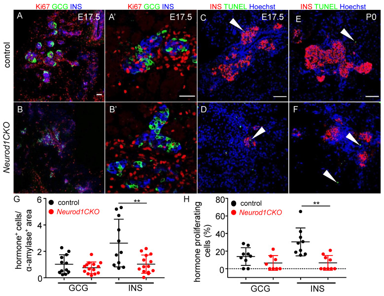
Diabetes is a metabolic disease that involves the death or dysfunction of the insulin-secreting β cells in the pancreas. Consequently, most diabetes research is aimed at understanding the molecular and cellular bases of pancreatic development, islet formation, β-cell survival, and insulin secretion. Complex interactions of signaling pathways and transcription factor networks regulate the specification, growth, and differentiation of cell types in the developing pancreas. Many of the same regulators continue to modulate gene expression and cell fate of the adult pancreas. The transcription factor NEUROD1 is essential for the maturation of β cells and the expansion of the pancreatic islet cell mass. Mutations of the Neurod1 gene cause diabetes in humans and mice. However, the different aspects of the requirement of NEUROD1 for pancreas development are not fully understood. In this study, we investigated the role of NEUROD1 during the primary and secondary transitions of mouse pancreas development. We determined that the elimination of Neurod1 impairs the expression of key transcription factors for α- and β-cell differentiation, β-cell proliferation, insulin production, and islets of Langerhans formation. These findings demonstrate that the Neurod1 deletion altered the properties of α and β endocrine cells, resulting in severe neonatal diabetes, and thus, NEUROD1 is required for proper activation of the transcriptional network and differentiation of functional α and β cells.

Keywords: NEUROD1; genetic mutation; mouse model; pancreatic development; transcriptional network.


Conflict of interest statement

The authors declare no conflict of interest.

Figures

Figure 1
Figure 1
Neurod1CKO mutants demonstrate a neonatal diabetic phenotype. (A) Breeding scheme shows genotypes for heterozygous (HET), homozygous mutant Neurod1CKO and control mice. The Isl1Cre transgene is transmitted paternally to eliminate any potential maternal influence on the developing embryos. (B) The survival of Neurod1CKO embryos is not affected, as shown by chi-squared test. (C) The blood glucose levels of control and Neurod1CKO pups fed ad libitum at P0 and P2-P3. Data are presented as mean ± SD, One-Way ANOVA (**** p < 0.0001).
Figure 2
Figure 2
NEUROD1 is efficiently reduced during the primary transition of early pancreas development in Neurod1CKO. (A) Representative whole-mount immunolabeling of dorsal pancreas shows reduction of NEUROD1+ clusters in the PDX1+ pancreatic domain (green) of Neurod1CKO compare to the control pancreas at E9.5 and E10.5. Isl1Cre expression is indicated by anti-Cre antibody (red). Large NEUROD1+ and Isl1-Cre+ cell clusters are detected in the control dorsal pancreas (arrowheads) but not in Neurod1CKO. The inverted single-channel image shows NEUROD1 expression. Asterisks indicate autofluorescent red blood cells. Scale bar: 50 μm. (B) Quantification of the NEUROD1+ area as a percentage of the total PDX1+ area in the dorsal pancreas at E10.5 using ImageJ program. Data are presented as mean ± SD, n = 4 embryos/genotype, Unpaired t-test (*** p = 0.0005).
Figure 3
Figure 3
The formation of glucagon+ clusters is unaffected but glucagon+ cells co-express PDX1 in Neurod1CKO during the primary transition. (A,B) Representative immunolabeling of the first endocrine cells expressing glucagon (GCG) is shown in the dorsal pancreatic epithelium delineated by the expression of PDX1 (whole-mounts). (A′,B′) Higher-magnification images show increased co-expression of PDX1 and GCG in Neurod1CKO compared to control pancreatic epithelium. (CE) Representative whole-mount immunolabeling shows dorsal and ventral pancreatic buds with GCG+ cells at E12.5. GCG+ area in the dorsal pancreas was quantified using ImageJ program and depicted as a percentage of GCG+ of the PDX1+ area in control and Neurod1CKO littermates (E). (F,G) Insulin (INS) expression in endocrine clusters is immunodetected in the control pancreas (arrowheads) but not in Neurod1CKO at E12.5 (vibratome sections). (H,I) NEUROG3 expressing endocrine precursors are shown in the whole-mount of control and Neurod1CKO PDX1+ dorsal pancreas. (J) NEUROG3+ cells were counted in the PDX1+ dorsal pancreas at E11.5. Data are presented as mean ± SD, Unpaired t-test (* p = 0.0354). Scale bars: 50 μm.
Figure 4
Figure 4
Reduced expression of genes important for the differentiation and function α and β cells in the Neurod1CKO pancreas at E14.5. Quantitative RT-PCR analyses of mRNA levels of transcription factors (A) and endocrine hormones (B) in E14.5 total pancreas of Neurod1CKO and control embryos. Genes highlighted in bold print indicate NEUROD1 target genes, as determined by ChIP-Seq analysis of NEUROD1 binding at mouse islet enhancers [31]. Data are presented as mean ± SEM (n = 8), Unpaired t-test (**** p < 0.0001, *** p < 0.001, ** p < 0.01, * p < 0.05). Ins1, insulin 1; Ins2, insulin 2; Gcg, glucagon; Sst, somatostatin; Ppy, pancreatic polypeptide; Ghrl, ghrelin.
Figure 5
Figure 5
Percentage of proliferating NEUROG3+ endocrine precursors is higher in the Neurod1CKO pancreas compared to the littermate controls. (A,B) Immunostaining for proliferating cell nuclear antigen Ki67 (red) in vibratome sections of the control and Neurod1CKO pancreas at E15.5 (arrows indicate NEUROG3+ proliferating cells). Nuclei are stained with Hoechst. (C) Quantification of proliferating NEUROG3+ cells per NEUROG3+ population (n = 3 embryos per genotype and 3–4 fields per section). Data are presented as mean ± SD, Unpaired t-test (* p = 0.03). Scale bar: 50 μm.
Figure 6
Figure 6
The formation of pancreatic islets is altered in the Neurod1CKO pancreas. Representative images of immunolabeling showing glucagon (GCG) producing α cells and β cells expressing PDX1, a marker of differentiated β cells, and insulin (INS) in the control and Neurod1CKO pancreas at E15.5 (A,B), and E17.5 (C,D). Higher-magnification images show a detail of the architecture of the islets of Langerhans at E15.5 (A′,B′), and E17.5 (C′,D′). Note decreased INS production, GCG+ cells co-expressing PDX1 (arrowhead), and the disrupted β-cell core/α-cell mantle organization of the islets in Neurod1CKO. Scale bars: 50 μm.
Figure 7
Figure 7
Proliferation potential of β cells is reduced in the Neurod1CKO pancreas. (A,B) Representative sections from the control and Neurod1CKO pancreas immunostained for proliferating cell nuclear antigen Ki67 (red) in endocrine α (glucagon, GCG) and β cells (insulin, INS) at E17.5. Note a disrupted formation of the islets of Langerhans in Neurod1CKO pancreas (A′,B′) Higher-magnification images show a detail of proliferating GCG+ and INS+ cells. (CF) Expression analysis of insulin (INS) and TUNEL staining performed on sections of E17.5 and P0 pancreata. Arrowheads indicate TUNEL+ cells. Nuclei are stained with Hoechst. (G) Relative quantification of GCG+ and INS+ cells per α-amylase+ area (marker of exocrine tissue) and (H) the percentage of GCG+ and INS+ cells expressing Ki67 per total number of GCG+ and INS+ cells in 80-μm vibratome sections of the pancreas at E17.5. Data are presented as mean ± SD; n = 3/genotype/3–5 fields of view, Unpaired t-test (** p = 0.0056 (G); ** p = 0.0011 (H)). Scale bars: 50 μm.
Figure 8
Figure 8
Insulin production is reduced in β cells and the differentiation of α cells is altered in the Neurod1CKO pancreas. (A,B) Representative vibratome sections from the control and Neurod1CKO pancreas show the expression of insulin (INS), glucagon (GCG) and PDX1, a marker of differentiated β cells. Note perturbations in the Neurod1CKO endocrine α cells co-expressing GCG and PDX1 (arrowheads), and β cells with PDX1 expressing cells without the expression of INS (arrows). (CF) Immunostaining for C-peptide 1 and C-peptide 2 shows a significant loss of C-peptide 1 levels in the Neurod1CKO pancreas. Nuclei are stained with Hoechst. (G,H) Reduced expression of C-peptide 1 is noticeable in the Neurod1CKO pancreas compared to littermate control at E17.5. Arrowheads indicate INS expressing cells without C-peptide 1 expression. (I) Quantitative RT-PCR analyses of mRNA levels of transcription factors and endocrine hormones in P1 total pancreas of Neurod1CKO and controls, indicating a major loss of β and α cells in the Neurod1CKO mutant. Data are presented as mean ± SEM (n = 8), Unpaired t-test (**** p < 0.0001, *** p < 0.001, ** p < 0.01). Ins1, insulin 1; Ins2, insulin 2; Gcg, glucagon. Scale bars: 50 μm.

Similar articles

Cited by

References

    1. Pan F.C., Wright C. Pancreas organogenesis: From bud to plexus to gland. Dev. Dyn. 2011;240:530–565. doi: 10.1002/dvdy.22584. - DOI - PubMed
    1. Bastidas-Ponce A., Scheibner K., Lickert H., Bakhti M. Cellular and molecular mechanisms coordinating pancreas development. Development. 2017;144:2873–2888. doi: 10.1242/dev.140756. - DOI - PubMed
    1. Gu G., Dubauskaite J., Melton D.A. Direct evidence for the pancreatic lineage: NGN3+ cells are islet progenitors and are distinct from duct progenitors. Development. 2002;129:2447–2457. doi: 10.1242/dev.129.10.2447. - DOI - PubMed
    1. Herrera P.L., Huarte J., Sanvito F., Meda P., Orci L., Vassalli J.D. Embryogenesis of the murine endocrine pancreas; early expression of pancreatic polypeptide gene. Development. 1991;113:1257–1265. doi: 10.1242/dev.113.4.1257. - DOI - PubMed
    1. Johansson K.A., Dursun U., Jordan N., Gu G., Beermann F., Gradwohl G., Grapin-Botton A. Temporal control of neurogenin3 activity in pancreas progenitors reveals competence windows for the generation of different endocrine cell types. Dev. Cell. 2007;12:457–465. doi: 10.1016/j.devcel.2007.02.010. - DOI - PubMed

MeSH terms

Substances

LinkOut - more resources