. 2021 Jun 23;22(13):6725.
doi: 10.3390/ijms22136725.

Membrane Interactions Accelerate the Self-Aggregation of Huntingtin Exon 1 Fragments in a Polyglutamine Length-Dependent Manner

Affiliations

Membrane Interactions Accelerate the Self-Aggregation of Huntingtin Exon 1 Fragments in a Polyglutamine Length-Dependent Manner

Arnaud Marquette et al. Int J Mol Sci. .

Abstract

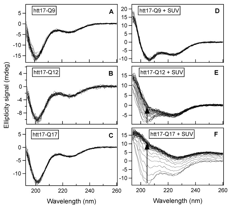
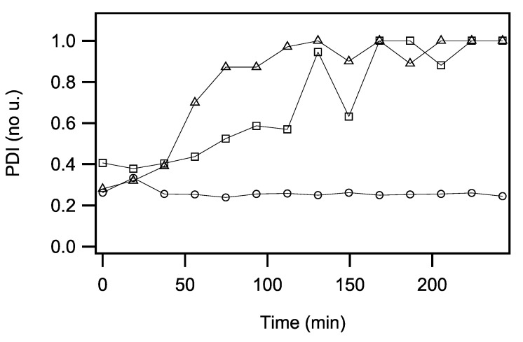
The accumulation of aggregated protein is a typical hallmark of many human neurodegenerative disorders, including polyglutamine-related diseases such as chorea Huntington. Misfolding of the amyloidogenic proteins gives rise to self-assembled complexes and fibres. The huntingtin protein is characterised by a segment of consecutive glutamines which, when exceeding ~ 37 residues, results in the occurrence of the disease. Furthermore, it has also been demonstrated that the 17-residue amino-terminal domain of the protein (htt17), located upstream of this polyglutamine tract, strongly correlates with aggregate formation and pathology. Here, we demonstrate that membrane interactions strongly accelerate the oligomerisation and β-amyloid fibril formation of htt17-polyglutamine segments. By using a combination of biophysical approaches, the kinetics of fibre formation is investigated and found to be strongly dependent on the presence of lipids, the length of the polyQ expansion, and the polypeptide-to-lipid ratio. Finally, the implications for therapeutic approaches are discussed.

Keywords: Huntington’s disease; amyloid; circular dichroism; dynamic light scattering; htt17; huntingtin; membrane-driven aggregation; peptide-lipid interactions; thioflavin T fluorescence.


Conflict of interest statement

The authors declare no conflict of interest.

Figures

Figure 1
Figure 1
Time-dependent structural changes measured by circular dichroism: CD spectra of htt17-Q9, htt17-Q12 and htt17-Q17 (C = 9.1 × 10⁻² mg/mL) in 10 mM Tris-HCl, pH 7 (AC), and in presence of SUVs made of POPC/POPS 3/1 mole/mole (C = 0.45 mg/mL) (DF) were recorded every 24.5 min. The progress of the spectral changes with time is depicted by arrows in panels E and F. Thereby the peptide-to-lipid ratios are 1/19.6, 1/22, and 1/26 for htt17-Q9, htt17-Q12, and htt17-Q17, respectively.
Figure 2
Figure 2
Aggregation kinetics by circular dichroism: the time-dependent intensity of the CD signal measured at 208 nm is shown for htt17-Q9, htt17-Q12, and htt17-Q17 in the presence of 0.45 mg/mL SUVs made of POPC/POPS 3/1 mole/mole in 10 mM Tris-HCl, pH 7. The results of least square fits with a mono-exponential function are displayed as solid lines. An error of ±1 was estimated from the signal-to-noise ratio of the spectra.
Figure 3
Figure 3
Time-dependent amyloid formation of htt17-Q17: the thioflavin T fluorescence is shown in the presence of 14.5 µM htt17-Q17 and SUVs made of 320 µM POPC/POPS 3/1 mole/mole in 10 mM Tris-HCl buffer, pH 7 (A). The peptide-to-lipid molar ratio was 1/22 and a spectrum was recorded every 2 min. The control experiments show almost no changes in thioflavin T fluorescence when exposed to the same amount of htt17-Q17 only (B) or SUVs only (C). The thioflavin T concentration was 5 µM in all recordings.
Figure 4
Figure 4
Time-dependent thioflavin T fluorescence as a function of polypeptide concentration: the thioflavin T fluorescence was continuously measured at 485 nm in the presence of SUVs and htt17-Q9 (A), htt17-Q12 (B), or htt17-Q17 (C), in 10 mM Tris-HCl, pH7. The peptide-to-lipid molar ratios are 1/22, 1/44, and 1/88, displayed as solid, short-dashed, and long-dashed lines, respectively. The lipid concentration was kept constant (C ≈ 320 µM) while the amount of peptide was adjusted to obtain the P/L ratios indicated. The ThT concentration was ≈ 5 µM in all the recordings.
Figure 5
Figure 5
Time-dependent changes of polydispersity: the polydispersity index (PDI) is as a function of time for SUVs in the presence of htt17-Q9 (circles), htt17-Q12 (squares), or htt17-Q17 (triangles) in 10 mM Tris-HCl, pH7. Measurements were performed by dynamic light scattering in the absence of mechanical stirring. The same concentrations of lipids and peptides were used as in the CD experiments presented in Figure 1 and Figure 2. Data points were recorded every 19–20 min.

Similar articles

Cited by

References

    1. Kuiper E.F., de Mattos E.P., Jardim L.B., Kampinga H.H., Bergink S. Chaperones in Polyglutamine Aggregation: Beyond the Q-Stretch. Front. Neurosci. 2017;11:145. doi: 10.3389/fnins.2017.00145. - DOI - PMC - PubMed
    1. Aktar F., Burudpakdee C., Polanco M., Pei S., Swayne T.C., Lipke P.N., Emtage L. The huntingtin inclusion is a dynamic phase-separated compartment. Life Sci. Alliance. 2019;2 doi: 10.26508/lsa.201900489. - DOI - PMC - PubMed
    1. Peskett T.R., Rau F., O’Driscoll J., Patani R., Lowe A.R., Saibil H.R. A Liquid to Solid Phase Transition Underlying Pathological Huntingtin Exon1 Aggregation. Mol. Cell. 2018;70:588–601.e586. doi: 10.1016/j.molcel.2018.04.007. - DOI - PMC - PubMed
    1. Pandey M., Rajamma U. Huntington’s disease: The coming of age. J. Genet. 2018;97:649–664. doi: 10.1007/s12041-018-0957-1. - DOI - PubMed
    1. Rubinsztein D.C., Leggo J., Coles R., Almqvist E., Biancalana V., Cassiman J.J., Chotai K., Connarty M., Crauford D., Curtis A., et al. Phenotypic characterization of individuals with 30-40 CAG repeats in the Huntington disease (HD) gene reveals HD cases with 36 repeats and apparently normal elderly individuals with 36-39 repeats. Am. J. Hum. Genet. 1996;59:16–22. - PMC - PubMed

LinkOut - more resources