Review
. 2021 Jun 23;22(13):6723.
doi: 10.3390/ijms22136723.

Usher Syndrome: Genetics of a Human Ciliopathy

Affiliations
Review

Usher Syndrome: Genetics of a Human Ciliopathy

Carla Fuster-García et al. Int J Mol Sci. .

Abstract

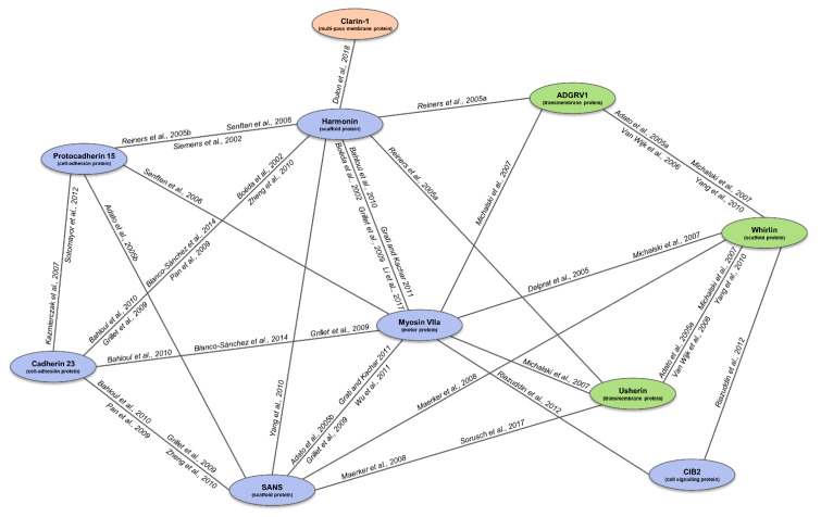
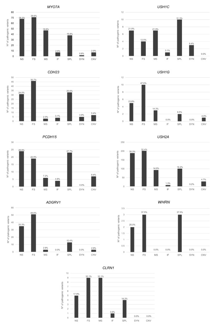
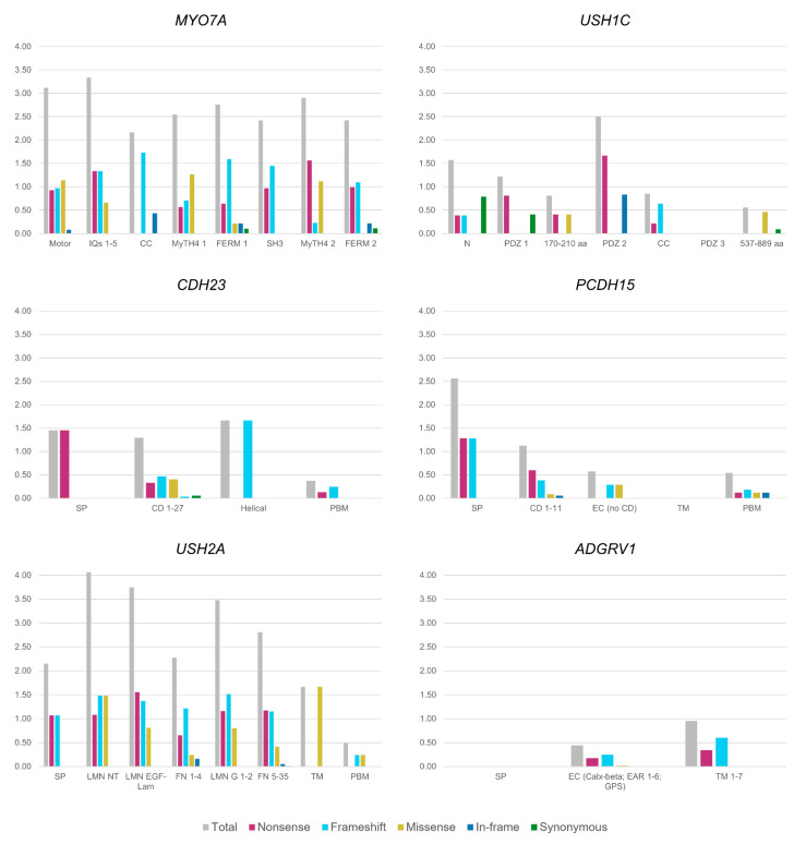
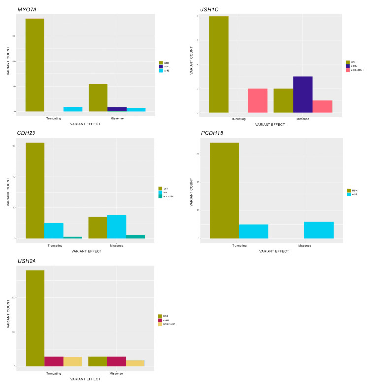
Usher syndrome (USH) is an autosomal recessive syndromic ciliopathy characterized by sensorineural hearing loss, retinitis pigmentosa and, sometimes, vestibular dysfunction. There are three clinical types depending on the severity and age of onset of the symptoms; in addition, ten genes are reported to be causative of USH, and six more related to the disease. These genes encode proteins of a diverse nature, which interact and form a dynamic protein network called the "Usher interactome". In the organ of Corti, the USH proteins are essential for the correct development and maintenance of the structure and cohesion of the stereocilia. In the retina, the USH protein network is principally located in the periciliary region of the photoreceptors, and plays an important role in the maintenance of the periciliary structure and the trafficking of molecules between the inner and the outer segments of photoreceptors. Even though some genes are clearly involved in the syndrome, others are controversial. Moreover, expression of some USH genes has been detected in other tissues, which could explain their involvement in additional mild comorbidities. In this paper, we review the genetics of Usher syndrome and the spectrum of mutations in USH genes. The aim is to identify possible mutation associations with the disease and provide an updated genotype-phenotype correlation.

Keywords: deafblindness; inherited retinal dystrophy; inner ear; pathogenic variant; photoreceptor; retinitis pigmentosa; sensorineural hearing loss; variant curation.


Conflict of interest statement

The authors declare that the research was conducted in the absence of any commercial or financial relationships that could be construed as a potential conflict of interest.

Figures

Figure 1
Figure 1
Representation of the USH interactome and supporting literature. The connecting lines represent the detected interactions between the USH1 (blue), USH2 (green) and USH3 (orange) proteins, according to the in vitro and in vivo studies to date (specified next to the lines). Adapted figure from Fuster García (2020). References figure: [10,92,106,107,108,138,139,140,141,142] [25,26,28,29,143,144,145] [61,146,147,148].
Figure 2
Figure 2
Mutation count in USH genes (MYO7A, USH1C, CDH23, PCDH15, USH2A, ADGRV1). Abbreviations: NS, nonsense variant; FS, frameshift variant; MS, missense variant; IF, in-frame indel variant; SYN, synonymous; SPL, splicing variant.
Figure 3
Figure 3
Mutational ratio in USH protein domains. Mutational ratio for the protein domains for the genes MYO7A, USH1C, CDH23, PCDH15, USH2A and ADGRV1, shown on the y-axis. Ratios are calculated counting the number of pathogenic variants of each type located in each domain and normalized with respect to the size of the specific domain. Abbreviations: Motor, motor domain; IQ, IQ calmodulin-binding motif; CC, coil-coiled domain; MyTH4; Myosin Tail Homology 4 domain; FERM, FERM domain (F for 4.1 protein, E for ezrin, R for radixin and M for moesin); SH3; Src Homology 3 domain; N, N-terminal domain; PDZ, PDZ domain; aa, amino acids; SP, signal peptide; PBM, PDZ binding domain; CD, cadherin domains; EC, extracellular domain; TM, transmembrane; LMN NT, laminin N-terminal domain; LMN EGF-Lam, laminin EGF-like domain; LMN G; laminin G domain; FN, fibronectin domain.
Figure 4
Figure 4
Mutational spectrum per gene for each phenotype. Abbreviations: USH, Usher syndrome; arHL, autosomal recessive hearing loss; adHL, autosomal dominant hearing loss; nsRP, non-syndromic retinitis pigmentosa. ADGRV1 is not included in the representation because all mutations obtained from the database were responsible for only USH and not for additional phenotypes.

Similar articles

Cited by

References

    1. Millán J.M., Aller E., Jaijo T., Blanco-Kelly F., Gimenez-Pardo A., Ayuso C. An Update on the Genetics of Usher Syndrome. J. Ophthalmol. 2011;2011:1–8. doi: 10.1155/2011/417217. - DOI - PMC - PubMed
    1. Wafa T.T., Faridi R., King K.A., Zalewski C., Yousaf R., Schultz J.M., Morell R.J., Muskett J., Turriff A., Tsilou E., et al. Vestibular phenotype-genotype correlation in a cohort of 90 patients with Usher syndrome. Clin. Genet. 2021;99:226–235. doi: 10.1111/cge.13868. - DOI - PMC - PubMed
    1. Davenport S.L.H., Omenn G.S. The Heterogeneity of Usher Syndrome; Proceedings of the 5th International Conference of Birth Defects; Montreal, QC, Canada. 21–27 August 1977.
    1. Weil D., Küssel P., Blanchard S., Levy G.G., Levi-Acobas F., Drira M., Ayadi H., Petit C. The autosomal recessive isolated deafness, DFNB2, and the Usher 1B syndrome are allelic defects of the myosin-VIIA gene. Nat. Genet. 1997;16:191–193. doi: 10.1038/ng0697-191. - DOI - PubMed
    1. Bork J.M., Peters L., Riazuddin S., Bernstein S.L., Ahmed Z.M., Ness S.L., Polomeno R., Ramesh A., Schloss M., Srisailpathy C.R.S., et al. Usher Syndrome 1D and Nonsyndromic Autosomal Recessive Deafness DFNB12 Are Caused by Allelic Mutations of the Novel Cadherin-Like Gene CDH23. Am. J. Hum. Genet. 2001;68:26–37. doi: 10.1086/316954. - DOI - PMC - PubMed