. 2021 Jun 23;22(13):6739.
doi: 10.3390/ijms22136739.

Treatment with the Bacterial Toxin CNF1 Selectively Rescues Cognitive and Brain Mitochondrial Deficits in a Female Mouse Model of Rett Syndrome Carrying a MeCP2-Null Mutation

Affiliations

Treatment with the Bacterial Toxin CNF1 Selectively Rescues Cognitive and Brain Mitochondrial Deficits in a Female Mouse Model of Rett Syndrome Carrying a MeCP2-Null Mutation

Chiara Urbinati et al. Int J Mol Sci. .

Abstract

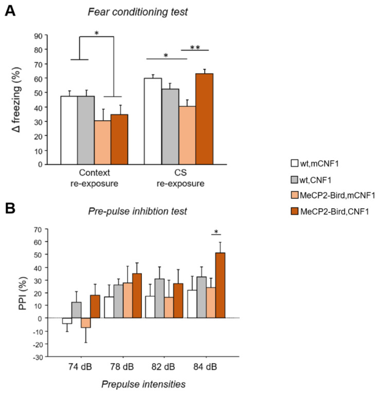
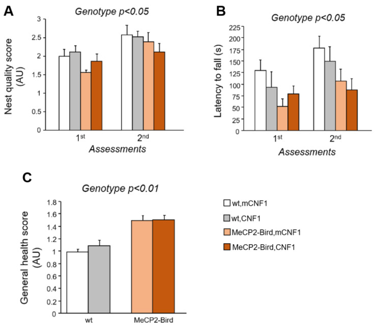
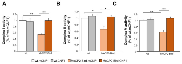
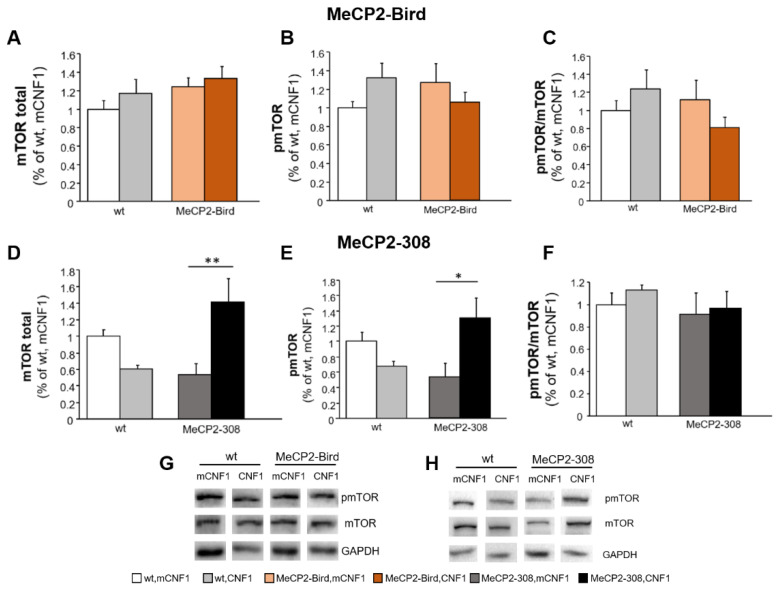
Rett syndrome (RTT) is a rare neurological disorder caused by mutations in the X-linked MECP2 gene and a major cause of intellectual disability in females. No cure exists for RTT. We previously reported that the behavioural phenotype and brain mitochondria dysfunction are widely rescued by a single intracerebroventricular injection of the bacterial toxin CNF1 in a RTT mouse model carrying a truncating mutation of the MeCP2 gene (MeCP2-308 mice). Given the heterogeneity of MECP2 mutations in RTT patients, we tested the CNF1 therapeutic efficacy in a mouse model carrying a null mutation (MeCP2-Bird mice). CNF1 selectively rescued cognitive defects, without improving other RTT-related behavioural alterations, and restored brain mitochondrial respiratory chain complex activity in MeCP2-Bird mice. To shed light on the molecular mechanisms underlying the differential CNF1 effects on the behavioural phenotype, we compared treatment effects on relevant signalling cascades in the brain of the two RTT models. CNF1 provided a significant boost of the mTOR activation in MeCP2-308 hippocampus, which was not observed in the MeCP2-Bird model, possibly explaining the differential effects of CNF1. These results demonstrate that CNF1 efficacy depends on the mutation beared by MeCP2-mutated mice, stressing the need of testing potential therapeutic approaches across RTT models.

Keywords: Rett syndrome; Rho GTPases; behaviour; cognition; energy metabolism; mTOR; mitochondria; mouse models.


Conflict of interest statement

The authors declare no conflict of interest.

Figures

Figure 1
Figure 1
CNF1 treatment improves MeCP2-Bird cued fear memory deficits and increases pre-pulse inhibition in MeCP2-Bird mice. (A) Contextual and cued fear memory were evaluated by testing experimental mice in a fear conditioning task. The Δ freezing was calculated as an index of fear memory and obtained by subtracting the percent time spent freezing during baseline from percent time spent freezing during either context or conditioned acoustic stimulus (CS) re-exposure. Symptomatic MeCP2-Bird female mice treated with an inactive form of the bacterial toxin (mCNF1) showed impaired memory for the aversive context and the CS, as demonstrated by lower Δ freezing showed during context and CS re-exposure, compared to wild type controls (wt, mCNF1). Treatment with CNF1 restored wt-like levels of Δ freezing to the CS, without affecting memory for the aversive context. (B) Experimental animals’ sensorimotor gating abilities were evaluated exposing mice to the pre-pulse inhibition (PPI) paradigm, consisting in reduction in the startle response to an acoustic pulse that occurs when presented with a pre-pulse stimulus. MeCP2-Bird mice did not display PPI deficits. Treatment with CNF1 tended to increase the PPI reflex in both wt and MeCP2-Bird mice, reaching a significant effect only in MeCP2-Bird mice at a pre-pulse intensity of 84 dB. The percentage of PPI was calculated, for each pre-pulse intensity, as follows: 100-[(mean startle amplitude for pre-pulse + pulse trials / mean startle amplitude for pulse alone trials) × 100]. Mice for each condition were as follows: wt, mCNF1: 7; wt, CNF1: 7; MeCP2-Bird, mCNF1: 6; MeCP2-Bird, CNF1: 6. Data are mean ± SEM. Statistical significance was assessed using three-way mixed ANOVA and Tukey’s post hoc tests. *: p < 0.05; **: p < 0.01.
Figure 2
Figure 2
Treatment with CNF1 does not rescue the motor deficits and the impaired general health status of symptomatic MeCP2-Bird female mice. (A) Coordinated and purposeful forepaw use was evaluated twice throughout the experimental schedule by the means of the nest building task. Nest quality was assessed by a trained observer blind to mouse genotype and treatment and measured in average units (AU). Symptomatic MeCP2-Bird female mice treated with an inactive form of the bacterial toxin (mCNF1) showed impaired nest building capacity relative to wild type (wt) controls, as demonstrated by the lower nest quality score. CNF1 treatment did not affect their performance. (B) Motor learning and coordination was evaluated through the accelerating rotarod task, that was repeated two times during the experiment. Symptomatic MeCP2-Bird females showed reduced motor coordination abilities, as represented by the lower latencies displayed to fall from the rotating rod. The graph represents the average latency out of three consecutive trials/assessment. (C) The general health status was evaluated throughout the experimental schedule by a trained observer blind to animals’ genotype and treatment. The average score of the five assessments is measured in AU and represented in the figure. Symptomatic MeCP2-Bird mice displayed impaired general health conditions compared to wt controls. Treatment with CNF1 was not able to rescue any of these behavioural impairments. Mice for each condition were as follows: wt, mCNF1: 7; wt, CNF1: 7; MeCP2-Bird, mCNF1: 5–6; MeCP2-Bird, CNF1: 6. Data are mean ± SEM. Statistical significance was assessed using three-way mixed ANOVA.
Figure 3
Figure 3
Treatment with CNF1 normalises the activity of mitochondrial respiratory chain complexes in symptomatic MeCP2-Bird mouse cortices. The activity of the mitochondrial respiratory chain (MRC) complexes I (A), II (B) and V (C) were measured spectrophotometrically in mitochondrial membrane-enriched fractions of experimental animals’ brain cortices. Symptomatic MeCP2-Bird female mice treated with an inactive form of the bacterial toxin (mCNF1) displayed reduced MRC complexes activity relative to wild type controls (wt, mCNF1). CNF1 restored wt-like levels of activity in treated MeCP2-Bird mice. Complex activities are expressed as percentage of the activity measured in wt, mCNF1 (set equal to one). Mice for each condition were as follows: wt, mCNF1: 3; wt, CNF1: 4; MeCP2-Bird, mCNF1: 4; MeCP2-Bird, CNF1: 4. Data are mean ± SEM obtained from three independent experiments. Statistical significance was assessed using two-way ANOVA and Tukey’s post hoc tests. *: p < 0.05; **: p < 0.01.
Figure 4
Figure 4
CNF1 treatment activates mTOR in the hippocampus of MeCP2-308 mice, but not in the MeCP2-Bird model. Total and phosphorylated (active) mTOR levels were evaluated by the means of Western blot analyses in hippocampi dissected from the brains of MeCP2-Bird, MeCP2-308 mice, and wild type (wt) littermates, treated with the recombinant (inactive, mCNF1) or active form of the bacterial toxin CNF1. (AC) mTOR total or phosphorylated (pmTOR) levels and pmTOR/mTOR ratio did not significantly differ among groups in the MeCP2-Bird cohort. (DF) CNF1 significantly increased both total and pmTOR levels in MeCP2-308 mice hippocampi but not the pmTOR/mTOR ratio. mTOR and pmTOR levels are normalised to total glyceraldehyde-3-phosphate dehydrogenase (GAPDH) contents and expressed as a proportion of those of wt, mCNF1 mice. (G,H) Immunoblots are examples from one animal of each experimental group. Mice for each condition were as follows: wt, mCNF1: 4 and 12; wt, CNF1: 4 and 12; MeCP2-Bird, mCNF1: 12; MeCP2-Bird, CNF1: 12; MeCP2-308, mCNF1: 4; MeCP2-308, CNF1: 4. Data are mean ± SEM. Statistical significance was assessed using two-way ANOVA and Tukey’s post hoc tests. *: p < 0.05; **: p < 0.01.

Similar articles

Cited by

References

    1. Chahrour M., Zoghbi H.Y. The story of Rett syndrome: From clinic to neurobiology. Neuron. 2007;56:422–437. doi: 10.1016/j.neuron.2007.10.001. - DOI - PubMed
    1. Ip J.P.K., Mellios N., Sur M. Rett syndrome: Insights into genetic, molecular and circuit mechanisms. Nat. Rev. Neurosci. 2018;19:368–382. doi: 10.1038/s41583-018-0006-3. - DOI - PMC - PubMed
    1. Shulyakova N., Andreazza A.C., Mills L.R., Eubanks J.H. Mitochondrial Dysfunction in the Pathogenesis of Rett Syndrome: Implications for Mitochondria-Targeted Therapies. Front. Cell. Neurosci. 2017;11:58. doi: 10.3389/fncel.2017.00058. - DOI - PMC - PubMed
    1. Valenti D., de Bari L., De Filippis B., Henrion-Caude A., Vacca R.A. Mitochondrial dysfunction as a central actor in intellectual disability-related diseases: An overview of Down syndrome, autism, Fragile X and Rett syndrome. Neurosci. Biobehav. Rev. 2014;46:202–217. doi: 10.1016/j.neubiorev.2014.01.012. - DOI - PubMed
    1. Ricceri L., De Filippis B., Laviola G. Rett syndrome treatment in mouse models: Searching for effective targets and strategies. Neuropharmacology. 2013;68:106–115. doi: 10.1016/j.neuropharm.2012.08.010. - DOI - PubMed

MeSH terms