. 2021 Jun 23;22(13):6742.
doi: 10.3390/ijms22136742.

The Effect of DREADD Activation of Leptin Receptor Positive Neurons in the Nucleus of the Solitary Tract on Sleep Disordered Breathing

Affiliations

The Effect of DREADD Activation of Leptin Receptor Positive Neurons in the Nucleus of the Solitary Tract on Sleep Disordered Breathing

Mateus R Amorim et al. Int J Mol Sci. .

Abstract

Obstructive sleep apnea (OSA) is recurrent obstruction of the upper airway due to the loss of upper airway muscle tone during sleep. OSA is highly prevalent, especially in obesity. There is no pharmacotherapy for OSA. Previous studies have demonstrated the role of leptin, an adipose-tissue-produced hormone, as a potent respiratory stimulant. Leptin signaling via a long functional isoform of leptin receptor, LEPRb, in the nucleus of the solitary tract (NTS), has been implicated in control of breathing. We hypothesized that leptin acts on LEPRb positive neurons in the NTS to increase ventilation and maintain upper airway patency during sleep in obese mice. We expressed designer receptors exclusively activated by designer drugs (DREADD) selectively in the LEPRb positive neurons of the NTS of Leprb-Cre-GFP mice with diet-induced obesity (DIO) and examined the effect of DREADD ligand, J60, on tongue muscle activity and breathing during sleep. J60 was a potent activator of LEPRb positive NTS neurons, but did not stimulate breathing or upper airway muscles during NREM and REM sleep. We conclude that, in DIO mice, the stimulating effects of leptin on breathing during sleep are independent of LEPRb signaling in the NTS.

Keywords: chemogenetics; obstructive sleep apnea; upper airway dysfunction.


Conflict of interest statement

The authors declare no conflict of interest.

Figures
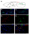
Figure 1
Figure 1
(A) Experimental design and injection sites of AAV8-hSyn-DIO-hM3D(Gq)-mCherry in the nucleus of the solitary tract (NTS) region. Localization of LEPRb positive neurons (green) in the NTS (B). The same, at a higher power (C). Transfection of DREADD (red in (D)) in the LEPRb positive neurons of the (green in (E)); DAPI (4′,6-diamidino-2-phenylindole) represents nuclei (blue in (F)). The orange color resulting from the red and green colors overlapping demonstrates DREADD expression in the LEPRb positive neurons (G). Scale bars, 50 µm. Arrows represent the co-localization of DREADD and GFP.
Figure 2
Figure 2
In-vitro activation of NTS neurons that contain DREADD in LEPRb positive neurons. (A) Representative tracing demonstrates an increase in action potential firing of the DREADD-containing NTS neuron, recorded in current-clamp configuration before and after J60 application. (B) The summary data from 5 neurons illustrate a significant increase in firing activity of NTS neurons post J60 application.
Figure 3
Figure 3
Sleep recordings. Representative traces of NREM sleep recordings in the same mouse expressing designer receptor exclusively activated by designer drug (DREADD) in the NTS after treatment with saline versus J60. * denotes breaths with inspiratory flow limitation.
Figure 4
Figure 4
Individual and grouped data showing the effects of J60 ligand or saline on maximal inspiratory flow (VImax), minute ventilation (VE), tidal volume (VT) and respiratory rate (RR) during flow-limited (A) and nonflow-limited breathing (B) in nonrapid eye movement (NREM) sleep.
Figure 5
Figure 5
Sleep recordings. Representative traces of REM sleep recordings in the same mouse expressing DREADD in the NTS after treatment with saline versus J60. * denotes breaths with inspiratory flow limitation.
Figure 6
Figure 6
Individual and grouped data showing the effects of J60 ligand or saline on maximal inspiratory flow (VImax), minute ventilation (VE), tidal volume (VT), and respiratory rate (RR) during flow-limited (A) and nonflow-limited breathing (B) in rapid eye movement (REM) sleep.
Figure 7
Figure 7
Genioglossus EMG recordings. (A) Representative genioglossal electromyography (EMGGG), moving average (ʃEMGGG), recorded at baseline (left) and after J60 administration (right) in a mouse expressing DREADD in the NTS. (B) Individual and grouped data showing the effects of J60 ligand on EMGGG in DREADD-treated animals.

Similar articles

Cited by

References

    1. Peppard P.E., Young T., Barnet J.H., Palta M., Hagen E.W., Hla K.M. Increased Prevalence of Sleep-Disordered Breathing in Adults. Am. J. Epidemiol. 2013;177:1006–1014. doi: 10.1093/aje/kws342. - DOI - PMC - PubMed
    1. Heinzer R., Vat S., Marques-Vidal P., Marti-Soler H., Andries D., Tobback N., Mooser V., Preisig M., Malhotra A., Waeber G., et al. Prevalence of Sleep-Disordered Breathing in the General Population: The HypnoLaus Study. Lancet Respir. Med. 2015;3:310–318. doi: 10.1016/S2213-2600(15)00043-0. - DOI - PMC - PubMed
    1. Young T., Palta M., Dempsey J., Skatrud J., Weber S., Badr S. The Occurrence of Sleep-Disordered Breathing among Middle-Aged Adults. N. Engl. J. Med. 1993;328:1230–1235. doi: 10.1056/NEJM199304293281704. - DOI - PubMed
    1. Tufik S., Santos-Silva R., Taddei J.A., Bittencourt L.R.A. Obstructive Sleep Apnea Syndrome in the Sao Paulo Epidemiologic Sleep Study. Sleep Med. 2010;11:441–446. doi: 10.1016/j.sleep.2009.10.005. - DOI - PubMed
    1. Punjabi N.M., Sorkin J.D., Katzel L.I., Goldberg A.P., Schwartz A.R., Smith P.L. Sleep-Disordered Breathing and Insulin Resistance in Middle-Aged and Overweight Men. Am. J. Respir. Crit. Care Med. 2002;165:677–682. doi: 10.1164/ajrccm.165.5.2104087. - DOI - PubMed