. 2021 Jun 23;22(13):6758.
doi: 10.3390/ijms22136758.

Hybrid Methacrylated Gelatin and Hyaluronic Acid Hydrogel Scaffolds. Preparation and Systematic Characterization for Prospective Tissue Engineering Applications

Affiliations

Hybrid Methacrylated Gelatin and Hyaluronic Acid Hydrogel Scaffolds. Preparation and Systematic Characterization for Prospective Tissue Engineering Applications

B Velasco-Rodriguez et al. Int J Mol Sci. .

Abstract

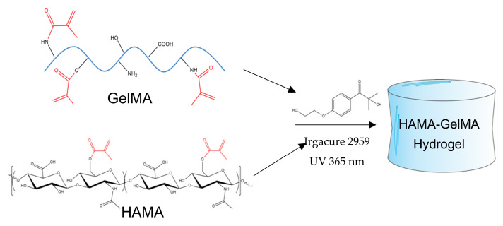
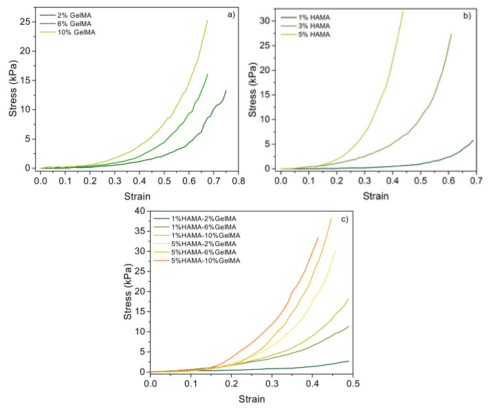
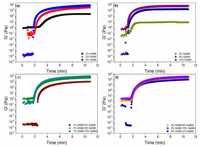
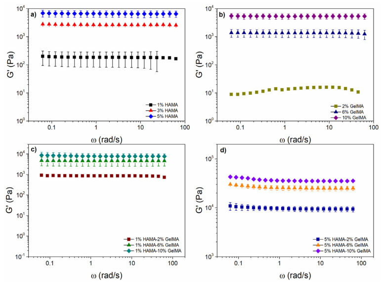
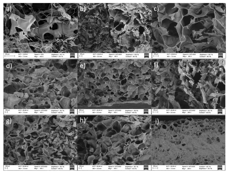
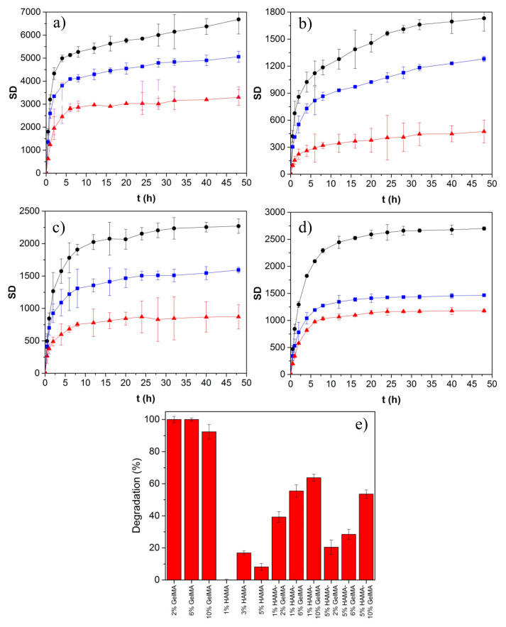
Hyaluronic acid (HA) and gelatin (Gel) are major components of the extracellular matrix of different tissues, and thus are largely appealing for the construction of hybrid hydrogels to combine the favorable characteristics of each biopolymer, such as the gel adhesiveness of Gel and the better mechanical strength of HA, respectively. However, despite previous studies conducted so far, the relationship between composition and scaffold structure and physico-chemical properties has not been completely and systematically established. In this work, pure and hybrid hydrogels of methacroyl-modified HA (HAMA) and Gel (GelMA) were prepared by UV photopolymerization and an extensive characterization was done to elucidate such correlations. Methacrylation degrees of ca. 40% and 11% for GelMA and HAMA, respectively, were obtained, which allows to improve the hydrogels' mechanical properties. Hybrid GelMA/HAMA hydrogels were stiffer, with elastic modulus up to ca. 30 kPa, and porous (up to 91%) compared with pure GelMA ones at similar GelMA concentrations thanks to the interaction between HAMA and GelMA chains in the polymeric matrix. The progressive presence of HAMA gave rise to scaffolds with more disorganized, stiffer, and less porous structures owing to the net increase of mass in the hydrogel compositions. HAMA also made hybrid hydrogels more swellable and resistant to collagenase biodegradation. Hence, the suitable choice of polymeric composition allows to regulate the hydrogels´ physical properties to look for the most optimal characteristics required for the intended tissue engineering application.

Keywords: gelatin; hyaluronic acid; hybrid scaffolds; hydrogel; porosity; tissue engineering.


Conflict of interest statement

The authors declare no conflict of interest.

Figures

Scheme A1
Scheme A1
Preparation of hybrid GelMA/HAMA hydrogels.
Figure A1
Figure A1
TGA scans of (a) pure HA and HAMA and (b) pure Gel and GelMA, and (c) plots of TGA and DSC runs of hybrid hydrogels with compositions 2% GelMA/1% HAMA and 10% GelMA/5% HAMA for comparison.
Figure A2
Figure A2
Storage modulus (G’) obtained from oscillatory deformation sweep measured for GelMA, HAMA, and mixtures of HAMA–GelMA at 37 °C.
Figure A3
Figure A3
Storage and loss moduli as a function of HAMA and GelMA concentrations for pure (a) HAMA and GelMA and (b,c) hybrid hydrogels at 37 °C and a frequency of 6.28 rad/s. Dashed lines represents the limits of the G values for use of the hydrogel as scaffolds for different types of tissues [5]. The continuous lines are to guide the eye.
Figure A4
Figure A4
Compression tests for pure (a) GelMA and (b) HAMA scaffolds and for hybrid (c) GelMA–HAMA of different compositions. Concentrations are indicated in the plots.
Figure 1
Figure 1
NMR spectra of (a) Gel () and GelMA (—) and (b) HA () and HAMA (—). In (a) and (b), the dotted rectangles denote the presence of the methacryloyl peaks after successful modification. In (a), the grey rectangle at ca. 7.1–7.5 ppm denotes the phenylalanine peaks taken as reference, whereas that one drawn at 3.0–3.3 ppm corresponds to the decrease of lysine groups. In (b), the grey rectangle indicates the methyl groups of HA at ca. 2.0 ppm taken as reference and the appearance of methyl protons of methacrylate at ca. 1.9 ppm, respectively.
Figure 2
Figure 2
(a,b) FTIR spectra of selected pure HAMA, GelMA, and hybrid GelMA/HAMA hydrogels at different compositions. Surface topographical composition acquired by Raman confocal 3D imaging of (c) 2% GelMA (green)-1% HAMA (red) and (d) 5% GelMA (red)-1% HAMA (green) hybrid hydrogels. HAMA was detected using bands at ca. 1380 and 1410 cm−1, whereas GelMA was detected using a band of ca. 1455 cm−1. (e) In-depth Raman imaging of the 2% GelMA-1% HAMA hydrogel. The bright blue-greenish color on the hydrogel in depth denotes the perfect mixing of HAMA (green) and GelMA (blue).
Figure 3
Figure 3
TGA scans of (a) hybrid GelMA/HAMA hydrogel scaffolds at different concentrations; (b) first derivative plots of TGA data of different pure and hybrid hydrogels; (c) examples of DSC curves corresponding pure Gel, pure HA, pure GelMA, pure HAMA, and some hybrid GelMA/HAMA hydrogel scaffolds.
Figure 4
Figure 4
Storage modulus (G′) as a function of the crosslinking time for different concentrations of (a) HAMA; (b) GelMA; (c) 1%HAMA-X% GelMA; and (d) 5% HAMA-X% GelMA at 37 °C.
Figure 5
Figure 5
Storage modulus as a function of frequency and (a) HAMA concentration; (b) GelMA concentration; (c) 1%HAMA-X% GelMA; and (d) 5% HAMA-X% GelMA. Performed at a deformation within the linear viscoelastic region (LVR) and a temperature of 37 °C.
Figure 6
Figure 6
SEM images of (a) 1%, (b) 3%, and (c) 5% HAMA; (d) 2% and (e) 10% GelMA; (f) 1% HAMA-2% GelMA; (g) 5% HAMA-2% GelMA; (h) 1% HAMA-10% GelMA; and (i) 5% HAMA-10% GelMA.
Figure 7
Figure 7
Swelling kinetic data for (a) pure HAMA hydrogels of (●) 1, () 3, and () 5%; (b) pure GelMA ones of (●) 2, () 6, and () 10%; and hybrid HAMA-GelMA ones of (c) (●) 1–2, () 1–6, and () 1–10%; and (d) (●) 5–2, () 5–6, and () 5–10%, respectively. (e) Degradation of scaffolds by enzymatic attack after 24 h of incubation at 37 °C.

Similar articles

Cited by

References

    1. Lee K.Y., Mooney D.J. Hydrogels for Tissue Engineering. Chem. Rev. 2001;101:1869–1880. doi: 10.1021/cr000108x. - DOI - PubMed
    1. Gu L., Li T., Song X., Yang X., Li S., Chen L., Liu P., Gong X., Chen C., Sun L. Preparation and characterization of methacrylated gelatin/bacterial cellulose composite hydrogels for cartilage tissue engineering. Regen. Biomater. 2020;7:195–202. doi: 10.1093/rb/rbz050. - DOI - PMC - PubMed
    1. Motealleh A., Kehr N.S. Nanocomposite Hydrogels and Their Applications in Tissue Engineering. Adv. Healthc. Mater. 2017;6:938. doi: 10.1002/adhm.201600938. - DOI - PubMed
    1. Zhang F., He C., Cao L., Feng W., Wang H., Mo X., Wang J. Fabrication of gelatin-hyaluronic acid hybrid scaffolds with tunable porous structures for soft tissue engineering. Int. J. Biol. Macromol. 2011;48:474–481. doi: 10.1016/j.ijbiomac.2011.01.012. - DOI - PubMed
    1. Vanderhooft J.L., Alcoutlabi M., Magda J.J., Prestwich G.D. Rheological properties of cross-linked hyaluronan-gelatin hydrogels for tissue engineering. Macromol. Biosci. 2009;9:20–28. doi: 10.1002/mabi.200800141. - DOI - PMC - PubMed

LinkOut - more resources