Review
. 2021 Jun 23;11(7):206.
doi: 10.3390/bios11070206.

Dengue Detection: Advances in Diagnostic Tools from Conventional Technology to Point of Care

Affiliations
Review

Dengue Detection: Advances in Diagnostic Tools from Conventional Technology to Point of Care

Md Alamgir Kabir et al. Biosensors (Basel). .

Abstract

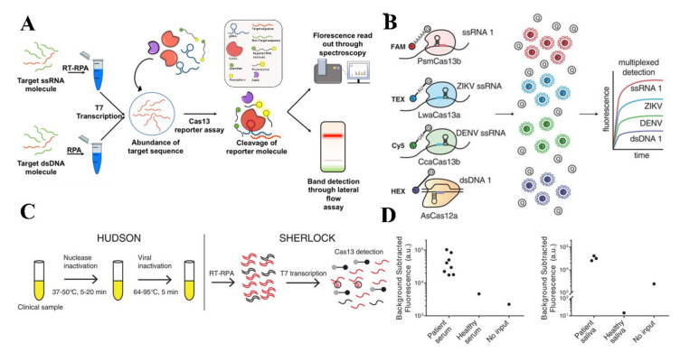
The dengue virus (DENV) is a vector-borne flavivirus that infects around 390 million individuals each year with 2.5 billion being in danger. Having access to testing is paramount in preventing future infections and receiving adequate treatment. Currently, there are numerous conventional methods for DENV testing, such as NS1 based antigen testing, IgM/IgG antibody testing, and Polymerase Chain Reaction (PCR). In addition, novel methods are emerging that can cut both cost and time. Such methods can be effective in rural and low-income areas throughout the world. In this paper, we discuss the structural evolution of the virus followed by a comprehensive review of current dengue detection strategies and methods that are being developed or commercialized. We also discuss the state of art biosensing technologies, evaluated their performance and outline strategies to address challenges posed by the disease. Further, we outline future guidelines for the improved usage of diagnostic tools during recurrence or future outbreaks of DENV.

Keywords: dengue; diagnostics; point-of-care.


Conflict of interest statement

Authors declare no financial conflict of interests.

Figures

Figure 1
Figure 1
(A) Worldwide estimated distribution of dengue in next 30 years due to the climatic and population change [8]. (B) Phylogeny analysis of 922 complete genomes evolution of all DENV serotypes reported from January 2000 to October 2020 based on their origin. (Source: Nextstrain.org (accessed on 15 May 2020)).
Figure 2
Figure 2
Structure of the dengue virus and evolution. (A) Dengue virus is spherical and about 40–60 nm in size. Genomic RNA and nucleocapsid proteins are covered by a membrane called Envelope. (B) The complete dengue virus genome is ~11 kb long, consisting of three structured and seven non-structured proteins and two untranslated regions (not drawn to the scale).
Figure 3
Figure 3
Immune response by the human body against the first invasion of the Dengue virus.
Figure 4
Figure 4
Detection of DENV RNA using isothermal amplification based platforms. (A) Detection of DENV RNA using LAMP method, initially RNA is transcripted to dsDNA and then amplified by the primers creating a loop structure (Modified from [107]). (B) A mobile laboratory to be used at point of care to detect DENV using RPA method utilizing minimal reagents as well as equipment (reprinted from [124]).
Figure 5
Figure 5
Detection of DENV viral biomarkers using surface plasmon resonance technologies. (A) An Au thin film based graphene oxide-polyamidoamine dendrimer based SPR sensor mechanism represents to measure the change in the SPR angle in present of DENV viral proteins (Reprinted from [128].). (B,C) Silver nanostructured SPR biosensor experimental setup to detect different concentration of NS1 antigen by measuring the peak wavelength shift on the chamber of a nanostructured device. NS1 antigen (green solid line), and anti-NS1 antibody (red dashed line) (Reprinted with permission from ref. [130]. Copyright 2019 Elsevier.). (D) Detection of different DENV serotypes based on altered distance based fluorescence signal (LSPR Enhancement and quenching) generated by primer–probe conjugated quantum dots and AuNPs (Reprinted from [133]). (E) AuNPs surface was modified by a self-assembled monolayer of MUA which facilitated covalent bonding of the DENV specific antibody with the AuNPs. (Reprinted with permission from ref. [134]. Copyright 2018 Elsevier.).
Figure 6
Figure 6
Electrochemical detection of DENV. (A) Electrochemical impedance sensing technique for DENV detection using graphene oxide and polymers (Reprinted with permission from ref. [154]. Copyright 2017 Elsevier.). (B) DENV virus detection using differential pulse voltammetric method utilizing a combination of nano composites (Reprinted with permission from ref. [160]. Copyright 2018 American Chemical Society.
Figure 7
Figure 7
Detection of DENV using microfluidic-based sensing technologies. (A) Experimental setup of a microfluidic chip based DENV detection using dielectrophoresis technique. AC voltage was supplied by a waveform generator and inverted microscope was used to record the fluorescence of the captured virus (Reprinted with permission from ref. [179]. Copyright 2017 Elsevier.) (B) A multilayered and PMMA made lab on a disc microfluidic device to ensure ultrasensitive detection of DENV (Reprinted from [185]). (C) Lab on a paper device to detect multiple pathogen targets accommodating amplification of RNA using LAMP method (Reprinted with permission from ref. [191]. Copyright 2020 Elsevier.).
Figure 8
Figure 8
(A)Detection of DENV using peptide microarrays. Differentiation of healthy and DENV infection patients’ sera by spotting the fluorescence (reprinted from [200]). (B) Detection of anti-DENV-1 antibody from whole blood using an intertwisted thread-based analytical device (Reprinted with permission from ref. [206]. Copyright 2020 American Chemical Society).
Figure 9
Figure 9
Detection of DENV using CRISPR-Cas based assays. (A) Graphical representation of SHERLOCK for detecting nucleic acids (reprinted from [215]). (B) Detection of in-sample four-channel multiplexed targets using SHERLOCK version 2 (Reprinted with permission from ref. [216]. Copyright 2018 The American Association for the Advancement of Science.). (C,D) The detection principle of clinical viral specimens from serum or saliva combining HUDSON and SHERLOCK (Reprinted with permission from ref. [217]. Copyright 2018 The American Association for the Advancement of Science.).

References

    1. Guzman M.G., Halstead S.B., Artsob H., Buchy P., Farrar J., Gubler D.J., Hunsperger E., Kroeger A., Margolis H.S., Martínez E. Dengue: A continuing global threat. Nat. Rev. Microbiol. 2010;8:S7–S16. doi: 10.1038/nrmicro2460. - DOI - PMC - PubMed
    1. Dick O.B., San Martín J.L., Montoya R.H., del Diego J., Zambrano B., Dayan G.H. The history of dengue outbreaks in the Americas. Am. J. Trop. Med. Hyg. 2012;87:584–593. doi: 10.4269/ajtmh.2012.11-0770. - DOI - PMC - PubMed
    1. World Health Organization . Dengue and Severe Dengue. World Health Organization, Regional Office for the Eastern Mediterranean; Cairo, Egypt: 2014.
    1. Bhatt S., Gething P.W., Brady O.J., Messina J.P., Farlow A.W., Moyes C.L., Drake J.M., Brownstein J.S., Hoen A.G., Sankoh O. The global distribution and burden of dengue. Nature. 2013;496:504–507. doi: 10.1038/nature12060. - DOI - PMC - PubMed
    1. Brady O.J., Gething P.W., Bhatt S., Messina J.P., Brownstein J.S., Hoen A.G., Moyes C.L., Farlow A.W., Scott T.W., Hay S.I. Refining the global spatial limits of dengue virus transmission by evidence-based consensus. PLoS Negl. Trop. Dis. 2012;6:e1760. doi: 10.1371/journal.pntd.0001760. - DOI - PMC - PubMed