Review
. 2021 Jun 25;10(13):2794.
doi: 10.3390/jcm10132794.

Systematic Review on Optical Diagnosis of Early Gastrointestinal Neoplasia

Affiliations
Review

Systematic Review on Optical Diagnosis of Early Gastrointestinal Neoplasia

Andrej Wagner et al. J Clin Med. .

Abstract

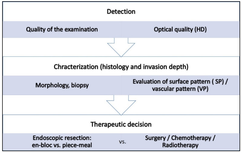
Background: Meticulous endoscopic characterization of gastrointestinal neoplasias (GN) is crucial to the clinical outcome. Hereby the indication and type of resection (endoscopically, en-bloc or piece-meal, or surgical resection) are determined. By means of established image-enhanced (IEE) and magnification endoscopy (ME) GN can be characterized in terms of malignancy and invasion depth. In this context, the statistical evidence and accuracy of these diagnostic procedures should be elucidated. Here, we present a systematic review of the literature.

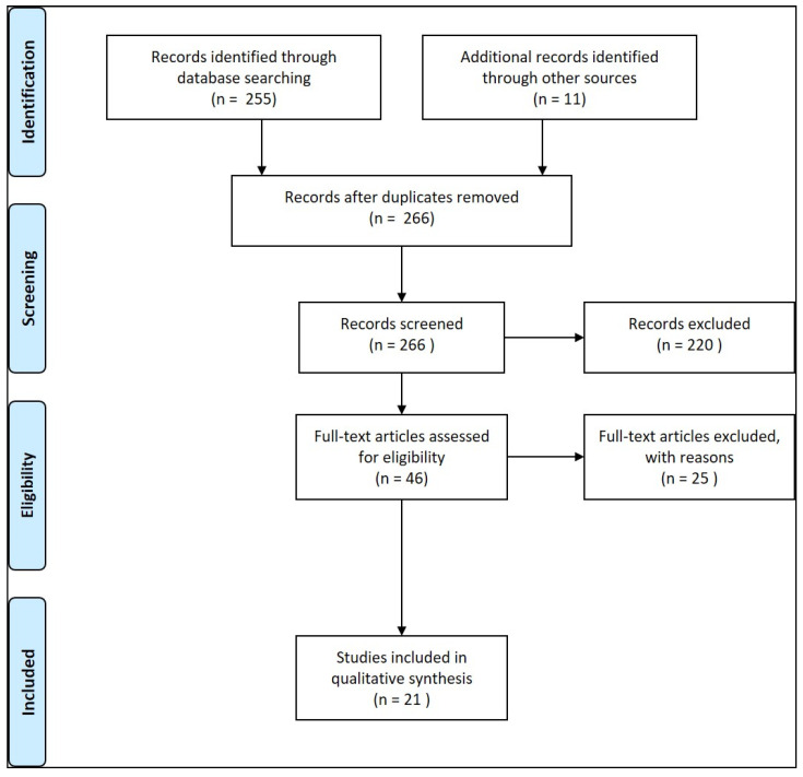
Results: 21 Studies could be found which met the inclusion criteria. In clinical prospective trials and meta-analyses, the diagnostic accuracy of >90% for characterization of malignant neoplasms could be documented, if ME with IEE was used in squamous cell esophageal cancer, stomach, or colonic GN.

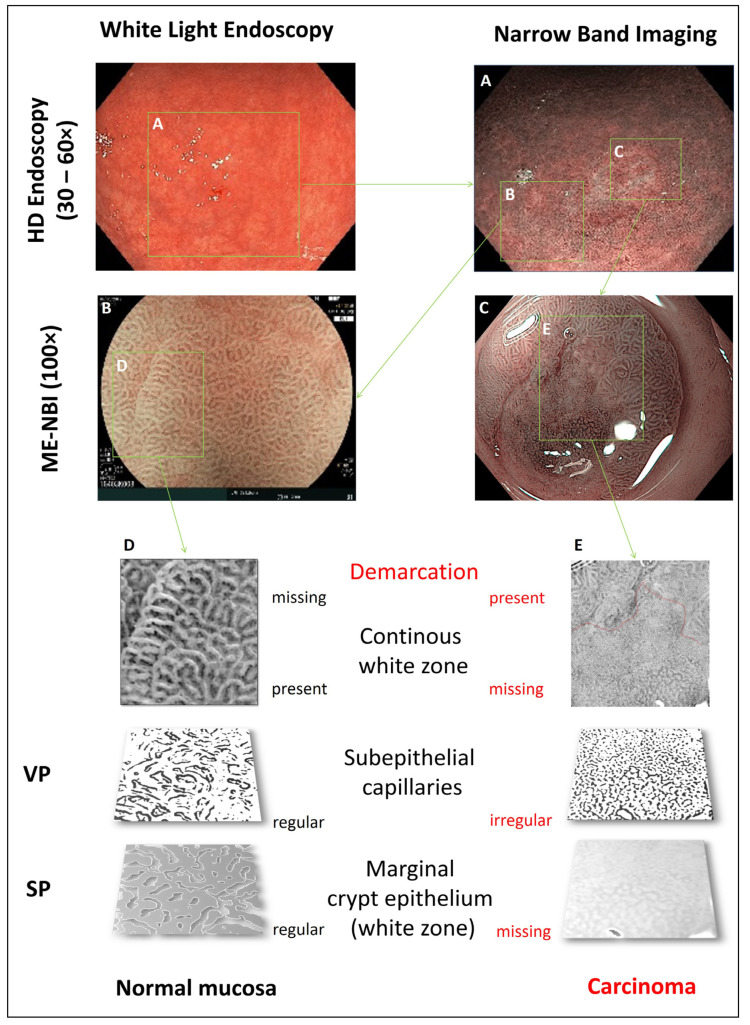
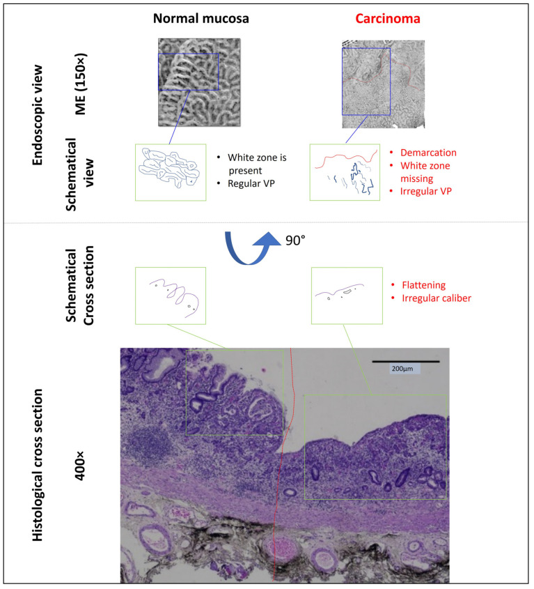
Conclusions: Currently, by means of optical diagnosis, today's gastrointestinal endoscopy is capable of determining the histological subtype, exact lateral spread, and depth of invasion of a lesion. The prerequisites for this are an exact knowledge of the anatomical structures, the endoscopic classifications based on them, and a systematic learning process, which can be supported by training courses. More prospective clinical studies are required, especially in the field of Barrett's esophagus and duodenal neoplasia.

Keywords: chromoendoscopy; endoscopy; invasion depth; magnification endoscopy; neoplasia.


Conflict of interest statement

The authors declare no conflict of interest.

Figures

Figure 1
Figure 1
The steps of the diagnostic process in endoscopy, the according key issues and their impact on therapeutic decisions.
Figure 2
Figure 2
Flow chart of the literature search.
Figure 3
Figure 3
Optical endoscopic characterization of a gastric lesion (antrum). (A): The lesion is merely demarcated in white light HD endoscopy. Magnification endoscopy (ME) with narrow band imaging (NBI) allows for characterization of normal vs. cancerous areas in the antrum (B,C). Determining factors are demarcation and presence of a white zone. Vascular pattern (VP) is determined by the pattern of subepithelial capillaries, and surface pattern (SP) by the marginal crypt epithelium (i.e., the “white zone”), respectively (D,E).
Figure 4
Figure 4
Comparison of magnification endoscopy view (ME) and histological cross section of normal mucosa and an early well differentiated adenocarcinoma in the antrum (compare Figure 2). The components of the ME picture, vascular pattern (VP) and surface pattern (SP), represent marginal crypt epithelium (i.e., the “white zone”) and subepithelial capillaries, respectively. SP and white zone are missing in the malignant area, VP is irregular. This corresponds to a flattening of the epithelium and irregular caliber of the capillary vessels in the histological specimen (obtained after endoscopic submucosal dissection of the lesion, which is characterized in Figure 2).
Figure 5
Figure 5
Endoscopic optical characterization of two large colonic lateral spreading tumors of the granular mixed type (LST gm). Magnification endoscopy with narrow band imaging (NBI) and chromoendoscopy with crystal violet allow for differentiation between early adenocarcinoma, which can be curatively resected by endoscopic submucosal dissection (A) and invasive carcinoma, requiring oncologic surgery (B).

References

    1. Ferlay J., Soerjomataram I., Dikshit R., Eser S., Mathers C., Rebelo M., Parkin D.M., Forman D., Bray F. Cancer incidence and mortality worldwide: Sources, methods and major patterns in globocan 2012. Int. J. Cancer. 2015;136:E359–E386. doi: 10.1002/ijc.29210. - DOI - PubMed
    1. Saitoh Y., Inaba Y., Sasaki T., Sugiyama R., Sukegawa R., Fujiya M. Management of colorectal t1 carcinoma treated by endoscopic resection. Dig. Endosc. 2016;28:324–329. doi: 10.1111/den.12503. - DOI - PubMed
    1. Ronnow C.F., Uedo N., Stenfors I., Toth E., Thorlacius H. Forceps biopsies are not reliable in the workup of large colorectal lesions referred for endoscopic resection: Should they be abandoned? Dis. Colon Rectum. 2019;62:1063–1070. doi: 10.1097/DCR.0000000000001440. - DOI - PubMed
    1. Vleugels J.L.A., Koens L., Dijkgraaf M.G.W., Houwen B., Hazewinkel Y., Fockens P., Dekker E. on behalf of the DISCOUNT study group. Suboptimal endoscopic cancer recognition in colorectal lesions in a national bowel screening programme. Gut. 2020;69:977–980. doi: 10.1136/gutjnl-2018-316882. - DOI - PMC - PubMed
    1. Atkinson N.S.S., Ket S., Bassett P., Aponte D., De Aguiar S., Gupta N., Horimatsu T., Ikematsu H., Inoue T., Kaltenbach T., et al. Narrow-band imaging for detection of neoplasia at colonoscopy: A meta-analysis of data from indi vidual patients in randomized controlled trials. Gastroenterology. 2019;157:462–471. doi: 10.1053/j.gastro.2019.04.014. - DOI - PubMed