. 2021 Jun 25;22(13):6827.
doi: 10.3390/ijms22136827.

Molecular and Electrophysiological Role of Diabetes-Associated Circulating Inflammatory Factors in Cardiac Arrhythmia Remodeling in a Metabolic-Induced Model of Type 2 Diabetic Rat

Affiliations

Molecular and Electrophysiological Role of Diabetes-Associated Circulating Inflammatory Factors in Cardiac Arrhythmia Remodeling in a Metabolic-Induced Model of Type 2 Diabetic Rat

Julian Zayas-Arrabal et al. Int J Mol Sci. .

Abstract

Background: Diabetic patients have prolonged cardiac repolarization and higher risk of arrhythmia. Besides, diabetes activates the innate immune system, resulting in higher levels of plasmatic cytokines, which are described to prolong ventricular repolarization.

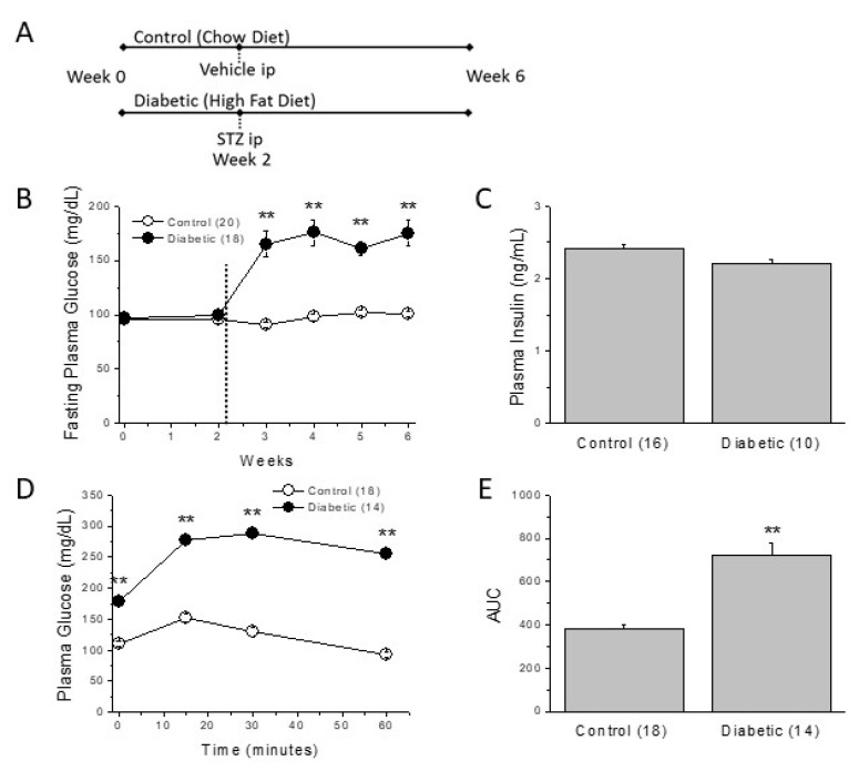
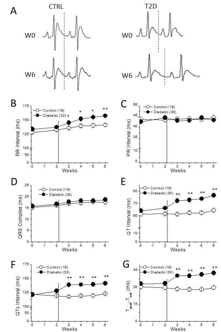
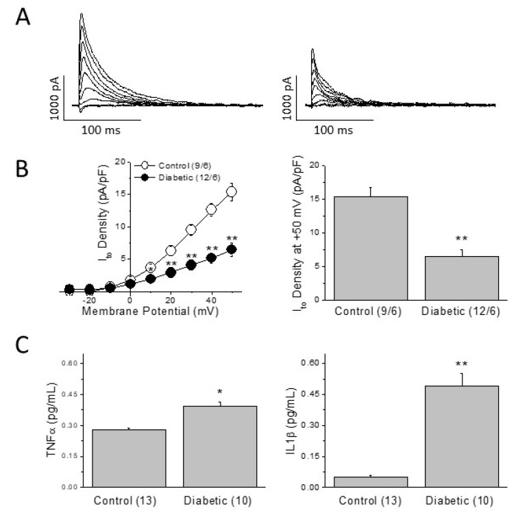
Methods: We characterize a metabolic model of type 2 diabetes (T2D) with prolonged cardiac repolarization. Sprague-Dawley rats were fed on a high-fat diet (45% Kcal from fat) for 6 weeks, and a low dose of streptozotozin intraperitoneally injected at week 2. Body weight and fasting blood glucose were measured and electrocardiograms of conscious animals were recorded weekly. Plasmatic lipid profile, insulin, cytokines, and arrhythmia susceptibility were determined at the end of the experimental period. Outward K+ currents and action potentials were recorded in isolated ventricular myocytes by patch-clamp.

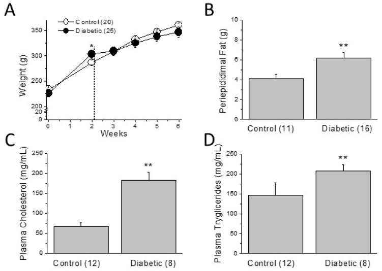
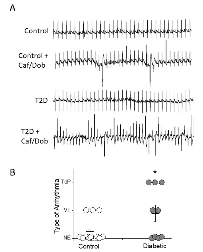
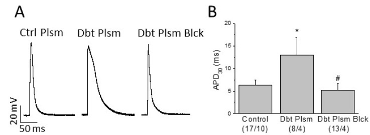
Results: T2D animals showed insulin resistance, hyperglycemia, and elevated levels of plasma cholesterol, triglycerides, TNFα, and IL-1b. They also developed bradycardia and prolonged QTc-interval duration that resulted in increased susceptibility to severe ventricular tachycardia under cardiac challenge. Action potential duration (APD) was prolonged in control cardiomyocytes incubated 24 h with plasma isolated from diabetic rats. However, adding TNFα and IL-1b receptor blockers to the serum of diabetic animals prevented the increased APD.

Conclusions: The elevation of the circulating levels of TNFα and IL-1b are responsible for impaired ventricular repolarization and higher susceptibility to cardiac arrhythmia in our metabolic model of T2D.

Keywords: arrhythmia; cytokines; insulin resistance; potassium current; torsade de pointes.


Conflict of interest statement

The authors declare no conflict of interest.

Figures

Figure 1
Figure 1
Type 2 diabetic rats show elevated plasma glucose and insulin resistance. (A) Description of the experimental protocol for induction of type 2 diabetes. (B) Weekly fasting plasma glucose throughout the experimental period. The dotted line indicates the injection of STZ or vehicle. (C) Plasma insulin levels after 4 weeks of diabetes. (D) IPIGTT performed at the end of the experimental period (week 6). (E) The corresponding area under the curve (AUC). In parenthesis (number of animals); ** p < 0.01.
Figure 2
Figure 2
Metabolic disturbances of the type 2 diabetic model. (A) Two weeks of HFD increased the body weight, but this effect was reversed by the STZ injection (dotted line). (B) Although total body weight is not different between groups at week 6, abdominal fat is significantly greater in diabetic animals. Plasma lipid profile worsened in diabetic animals as can be seen in the increased circulating levels of (C) cholesterol and (D) triglycerides. In parenthesis (number of animals); * p > 0.05; ** p < 0.01.
Figure 3
Figure 3
Cardiac electrical remodeling in diabetic heart. (A) Electrocardiographic recordings in a control and in a diabetic animal before and after the 6-weeks experimental period. The dotted line shows the end of the T wave. (BD) Cardiac impulse generation (RR interval) and conduction (PR interval and QRS complex) throughout the 6 weeks. (E,F) The main repolarization parameters (QT, QTc, and Tpeak-Tend) are longer in diabetic than in control animals. In parenthesis (number of animals); * p < 0.05; ** p < 0.01. The dotted line in (BG) shows the moment of STZ or vehicle injection.
Figure 4
Figure 4
Increased arrhythmia susceptibility in diabetic heart. (A) Examples of non-arrhythmic and arrhythmic electrocardiographic recordings in control and diabetic animals after caffeine/dobutamine challenge (Caf/Dob). The ECG of the control animal displays only ventricular premature beats and salvo, whereas the diabetic animal shows episodes of torsade de pointes (TdP). (B) Incidence and severity of ventricular tachycardias and TdP in control and diabetic animals. Each arrhythmia was scored as follows: No events (NE) = 0; ventricular tachycardia (VT) = 1; and TdP = 2. The incidence and severity of ventricular arrhythmias after cardiac challenge are higher in diabetic animals than in controls. * p < 0.05.
Figure 5
Figure 5
Repolarization capacity and inflammation. (A) Current traces of a current-voltage protocol of the transient outward current elicited in a control cell and a diabetic ventricular cell. (B) Ito density/voltage relationships and Ito density at +50 mV in control and diabetic myocytes. In parenthesis (number of cells/number of animals). (C) TNFα and IL-1b can reduce the density of Ito. Plasma levels of TNFα and IL-1b are higher in type 2 diabetic animals than in age-matched controls. Data are mean ± SEM; in parenthesis (number of animals); * p < 0.05; ** p < 0.01.
Figure 6
Figure 6
Role of circulating mediators on cardiac electrical remodeling. (A) Typical action potentials recorded in myocytes isolated from the right ventricle of healthy animals incubated for 24 h in DMEM supplemented with: plasma from healthy animals (Ctrl Plsm); with plasma from diabetic animals (Dbt Plsm); or plasma extracted from type 2 diabetic animals plus TNFα and IL-1b receptor blockers (Dbt Plsm Blck). (B) Incubation with diabetic plasma prolongs the action potential duration at 30% of repolarization (APD30), and this effect is prevented by TNFα plus IL-1b receptor blockers. Data are mean ± SEM; in parenthesis (number of cells/number of animals); * p < 0.05 with respect to control; # p < 0.05 with respect to Dbt Plsm.

Similar articles

Cited by

References

    1. El-Atat F.A., McFarlane S.I., Sowers J.R., Bigger J.T. Sudden cardiac death in patients with diabetes. Curr. Diabetes Rep. 2004;4:187–193. doi: 10.1007/s11892-004-0022-8. - DOI - PubMed
    1. Tse G., Lai E.T.H., Tse V., Yeo J.M. Molecular and electrophysiological mechanisms underlying cardiac arrhythmogenesis in diabetes mellitus. J. Diabetes Res. 2016;2016:2848759. doi: 10.1155/2016/2848759. - DOI - PMC - PubMed
    1. Sanguinetti M.C., Tristani-Firouzi M. hERG Potassium Channels and Cardiac Arrhythmia. Nature. 2006;440:463–469. doi: 10.1038/nature04710. - DOI - PubMed
    1. Kahn J.K., Sisson J.C., Vinik A.I. QT interval prolongation and sudden cardiac death in diabetic autonomic neuropathy. J. Clin. Endocrinol. Metab. 1987;64:751–754. doi: 10.1210/jcem-64-4-751. - DOI - PubMed
    1. Bellavere F., Ferri M., Guarini L., Bax G., Piccoli A., Cardone C., Fedele D. Prolonged QT period in diabetic autonomic neuropathy—a possible role in sudden cardiac death. Br. Heart J. 1988;59:379–383. doi: 10.1136/hrt.59.3.379. - DOI - PMC - PubMed