. 2021 Jun 25;11(7):1161.
doi: 10.3390/diagnostics11071161.

Dental Anomalies' Characteristics

Affiliations

Dental Anomalies' Characteristics

Tatiana Sella Tunis et al. Diagnostics (Basel). .

Abstract

The aim of this study was to characterize dental anomalies. The pretreatment records (photographs and radiographs) of 2897 patients (41.4% males and 58.6% females) were utilized to detect dental anomalies. The dental anomalies studied were related to number, size and shape, position, and eruption. A Chi-square test was carried out to detect associations between dental anomalies, jaw, and sex. A total of 1041 (36%) of the subjects manifested at least one dental anomaly. The prevalence of all dental anomalies was jaw-dependent and greater in the maxilla, except for submerged and transmigrated teeth. The most frequently missing teeth were the maxillary lateral incisor (62.3%) and the mandibular second premolars (60.6%). The most frequent supernumerary teeth were the incisors in the maxilla (97%) and the first premolars in the mandible (43%). Dental anomalies are more frequent in the maxilla and mainly involve the anterior teeth; in the mandible, however, it is the posterior teeth. These differences can be attributed to the evolutionary history of the jaws and their diverse development patterns.

Keywords: dental anomalies; dental diagnosis; growth and development; mandible; maxilla.


Conflict of interest statement

The authors declare no conflict of interest. The funders played no role in the design of the study; in the collection, analyses, or interpretation of data; in the writing of the manuscript; or in the decision to publish the results.

Figures
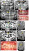
Figure 1
Figure 1
Examples of dental anomalies evaluated on radiographs and intraoral photographs: (a) panoramic radiograph of a patient with a submerged tooth #55 (yellow arrow); (b) panoramic radiograph of a patient with maxillary impacted canine teeth #23 and 13 (orange arrows), retained deciduous canine teeth #53 and 63 (green arrows), and a mandibular canine transmigration tooth #43 (yellow arrow) located at the lower border of the symphysis; (c) an example of a patient with a missing tooth #12 (yellow arrow) and a peg-shaped incisor tooth #22 (blue arrow): (c1) panoramic radiograph, (c2) intraoral frontal photograph, (c3) intraoral lateral photograph; (d) an example of a patient with a supernumerary tooth (impacted between the roots of the teeth #11–12, yellow arrow), impacted canine teeth #13 and 23 (blue arrows), and retained deciduous teeth #53 and 63 (green arrows): (d1) panoramic radiograph, (d2) periapical radiograph; (e) an example of a patient with dental transposition of the teeth #13 and #12 (yellow arrow): (e1) panoramic radiograph, (e2) periapical radiograph; (f) an example of a patient with fusion of the teeth #11 and 12 (yellow arrow): (f1) intraoral photograph, and (f2) periapical radiograph.
Figure 2
Figure 2
The relative frequencies of dental anomalies in the maxilla (blue) and mandible (red).
Figure 3
Figure 3
Distribution of dental anomalies by tooth type, jaw, and location (anterior (blue and green) vs. posterior (red and orange)): (a) missing teeth, (b) supernumerary teeth, (c) ectopic teeth, (d) impacted teeth, and (e) retained teeth.

References

    1. Fekonja A. Prevalence of dental developmental anomalies of permanent teeth in children and their influence on esthetics. J. Esthet. Restor. Dent. 2017;29:276–283. doi: 10.1111/jerd.12302. - DOI - PubMed
    1. Vastardis H., Karimbux N., Guthua S.W., Seidman J., Seidman C.E. A human MSX1 homeodomain missense mutation causes selective tooth agenesis. Nat. Genet. 1996;13:417–421. doi: 10.1038/ng0896-417. - DOI - PubMed
    1. Vitria E.E., Tofani I., Kusdhany L., Bachtiar E.W. Genotyping analysis of the Pax9 Gene in patients with maxillary canine impaction. F1000 Res. 2019;8:254. doi: 10.12688/f1000research.17147.1. - DOI - PMC - PubMed
    1. Azzaldeen A., Watted N., Mai A., Borbély P., Abu-Hussein M. Tooth agenesis; Aetiological factors. J. Dent. Med. Sci. 2017;16:75–85. doi: 10.9790/0853-1601057585. - DOI
    1. Shilpa G., Gokhale N., Mallineni S.K., Nuvvula S. Prevalence of dental anomalies in deciduous dentition and its association with succedaneous dentition: A cross-sectional study of 4180 South Indian children. J. Indian Soc. Pedod. Prev. Dent. 2017;35:56–62. doi: 10.4103/0970-4388.199228. - DOI - PubMed

LinkOut - more resources