. 2021 Jun 25;22(13):6837.
doi: 10.3390/ijms22136837.

Extracellular Vesicles Are More Potent Than Adipose Mesenchymal Stromal Cells to Exert an Anti-Fibrotic Effect in an In Vitro Model of Systemic Sclerosis

Affiliations

Extracellular Vesicles Are More Potent Than Adipose Mesenchymal Stromal Cells to Exert an Anti-Fibrotic Effect in an In Vitro Model of Systemic Sclerosis

Pauline Rozier et al. Int J Mol Sci. .

Abstract

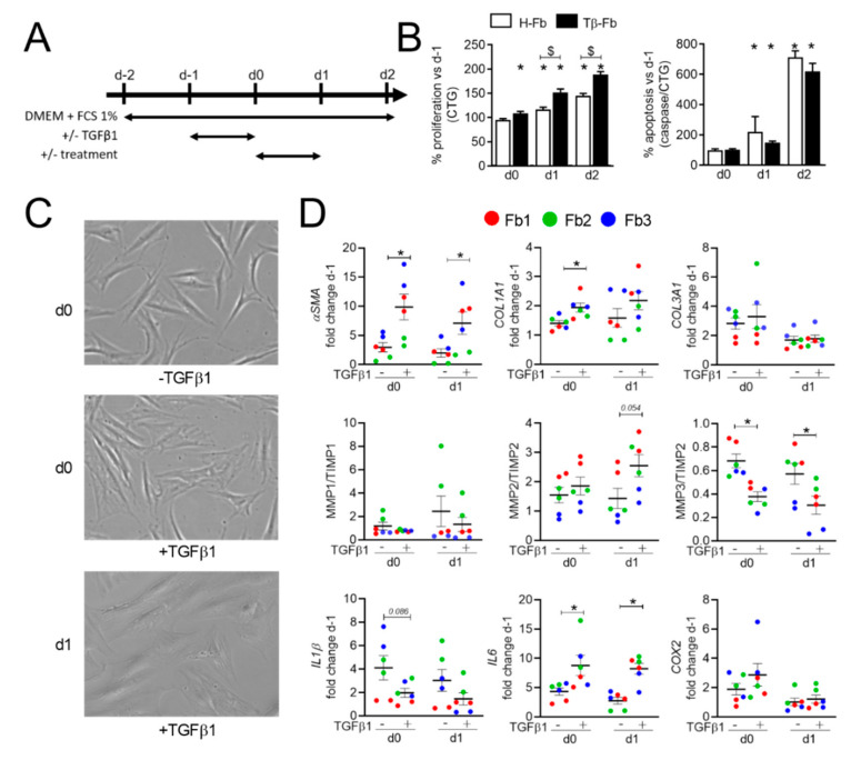
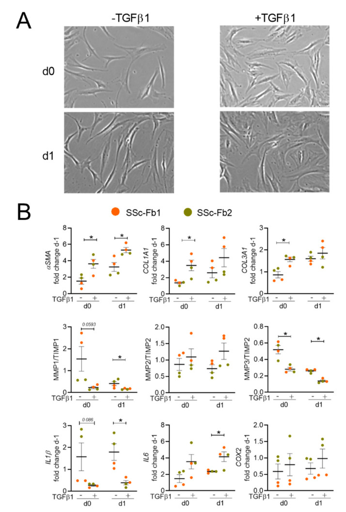
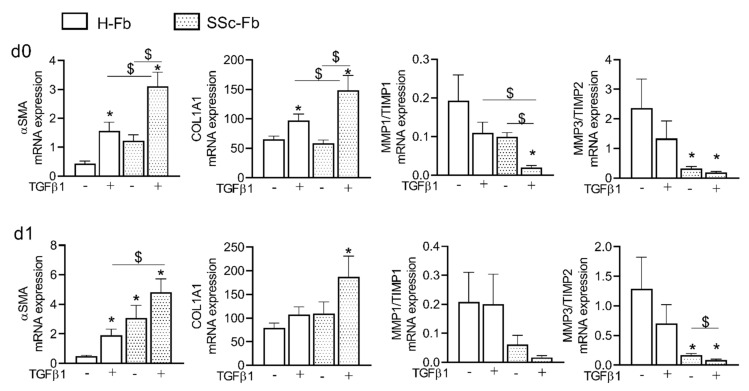
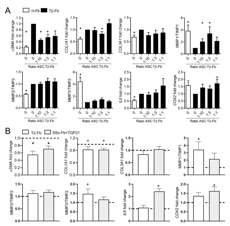
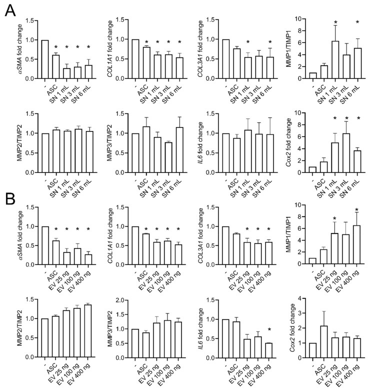
Systemic sclerosis (SSc) is a complex disorder resulting from dysregulated interactions between the three main pathophysiological axes: fibrosis, immune dysfunction, and vasculopathy, with no specific treatment available to date. Adipose tissue-derived mesenchymal stromal cells (ASCs) and their extracellular vesicles (EVs) have proved efficacy in pre-clinical murine models of SSc. However, their precise action mechanism is still not fully understood. Because of the lack of availability of fibroblasts isolated from SSc patients (SSc-Fb), our aim was to determine whether a TGFβ1-induced model of human myofibroblasts (Tβ-Fb) could reproduce the characteristics of SSc-Fb and be used to evaluate the anti-fibrotic function of ASCs and their EVs. We found out that Tβ-Fb displayed the main morphological and molecular features of SSc-Fb, including the enlarged hypertrophic morphology and expression of several markers associated with the myofibroblastic phenotype. Using this model, we showed that ASCs were able to regulate the expression of most myofibroblastic markers on Tβ-Fb and SSc-Fb, but only when pre-stimulated with TGFβ1. Of interest, ASC-derived EVs were more effective than parental cells for improving the myofibroblastic phenotype. In conclusion, we provided evidence that Tβ-Fb are a relevant model to mimic the main characteristics of SSc fibroblasts and investigate the mechanism of action of ASCs. We further reported that ASC-EVs are more effective than parental cells suggesting that the TGFβ1-induced pro-fibrotic environment may alter the function of ASCs.

Keywords: TGFβ1; anti-fibrotic; extracellular vesicles; mesenchymal stem cells; systemic sclerosis.


Conflict of interest statement

The authors disclose any financial or personal conflicts of interest.

Figures

Figure 1
Figure 1
In vitro model of Transforming Growth Factor β1 (TGFβ1)-stimulated fibroblasts. (A) Experimental scheme. Dermal fibroblasts from healthy donors (H-Fb) were starved in a DMEM medium containing 1% FCS (day-2) for 24 h before stimulation with TGFβ1 for 24 h. On day 0, treatment was applied for 24 h and samples were analyzed (day 1 or 2). (B) The proliferation of H-Fb or TGFβ1-stimulated fibroblasts (Tβ-Fb; left panel) normalized on day-1 (n = 12). Apoptosis normalized on CTG assay (right panel). (C) Representative pictures of H-Fb (−TGFβ1) on day 0 or Tβ-Fb (+TGFβ1) on days 0 and 1 after stimulation (×40 objective). (D) Fold change of gene expression normalized on day-1 (n = 6). *: p < 0.05 versus H-Fb on day 0 or $: p < 0.05 versus the indicated group.
Figure 2
Figure 2
The phenotype of fibroblasts isolated from systemic sclerosis (SSc) patients. (A) Representative pictures of fibroblasts from SSc patients (SSc-Fb) cultured with or without TGFβ1 on days 0 and 1 after stimulation (×40 objective). (B) Fold change of gene expression normalized on day-1 (n = 4). *: p < 0.05.
Figure 3
Figure 3
Comparison of the phenotype of healthy and SSc fibroblasts in standard culture conditions or after TGFβ1 stimulation. Fold change of gene expression (n = 12). *: p < 0.05 compared to H-Fb (−TGFβ1); $: p < 0.05 compared to the indicated group.
Figure 4
Figure 4
Effect of adipose tissue-derived mesenchymal stromal cells (ASCs) on TGFβ1-stimulated fibroblasts. (A) Fold change of gene expression in healthy fibroblasts (H-Fb) and TGFβ1-stimulated fibroblasts (Tβ-Fb) normalized to H-Fb (n = 14). (B) Fold change of gene expression of markers as in (A) in Tβ-Fb or TGFβ1-stimulated SSc-Fb (n = 8–14). *: p < 0.05.
Figure 5
Figure 5
Effects of ASC-EVs on TGFβ1-stimulated fibroblasts. (A) Fold change of gene expression in TGFβ1-stimulated fibroblasts using different volumes of conditioned supernatants (SN) from healthy ASCs (n = 4). (B) Fold change of gene expression as in (A) (n = 18). *: p < 0.05.

References

    1. Hughes L.C., Archer C.W., Ap Gwynn I. The ultrastructure of mouse articular cartilage: Collagen orientation and implications for tissue functionality. A polarised light and scanning electron microscope study and review. Eur. Cells Mater. 2005;9:68–84. doi: 10.22203/eCM.v009a09. - DOI - PubMed
    1. Varga J., Whitfield M.L. Transforming growth factor-beta in systemic sclerosis (scleroderma) Front. Biosci. 2009;1:226–235. doi: 10.2741/s22. - DOI - PubMed
    1. Lin S.J., Lerch T.F., Cook R.W., Jardetzky T.S., Woodruff T.K. The structural basis of TGF-beta, bone morphogenetic protein, and activin ligand binding. Reproduction. 2006;132:179–190. doi: 10.1530/rep.1.01072. - DOI - PubMed
    1. Massague J. TGF-beta signal transduction. Annu. Rev. Biochem. 1998;67:753–791. doi: 10.1146/annurev.biochem.67.1.753. - DOI - PubMed
    1. Milano A., Pendergrass S.A., Sargent J.L., George L.K., McCalmont T.H., Connolly M.K., Whitfield M.L. Molecular subsets in the gene expression signatures of scleroderma skin. PLoS ONE. 2008;3:e2696. doi: 10.1371/annotation/05bed72c-c6f6-4685-a732-02c78e5f66c2. - DOI - PMC - PubMed

Substances