Case Reports
. 2021 Jun 24;22(13):6775.
doi: 10.3390/ijms22136775.

The Double Mutation DSG2-p.S363X and TBX20-p.D278X Is Associated with Left Ventricular Non-Compaction Cardiomyopathy: Case Report

Affiliations
Case Reports

The Double Mutation DSG2-p.S363X and TBX20-p.D278X Is Associated with Left Ventricular Non-Compaction Cardiomyopathy: Case Report

Roman Myasnikov et al. Int J Mol Sci. .

Abstract

Left ventricular non-compaction cardiomyopathy (LVNC) is a rare heart disease, with or without left ventricular dysfunction, which is characterized by a two-layer structure of the myocardium and an increased number of trabeculae. The study of familial forms of LVNC is helpful for risk prediction and genetic counseling of relatives. Here, we present a family consisting of three members with LVNC. Using a next-generation sequencing approach a combination of two (likely) pathogenic nonsense mutations DSG2-p.S363X and TBX20-p.D278X was identified in all three patients. TBX20 encodes the cardiac T-box transcription factor 20. DSG2 encodes desmoglein-2, which is part of the cardiac desmosomes and belongs to the cadherin family. Since the identified nonsense variant (DSG2-p.S363X) is localized in the extracellular domain of DSG2, we performed in vitro cell transfection experiments. These experiments revealed the absence of truncated DSG2 at the plasma membrane, supporting the pathogenic relevance of DSG2-p.S363X. In conclusion, we suggest that in the future, these findings might be helpful for genetic screening and counseling of patients with LVNC.

Keywords: DSG2; TBX20; cardiomyopathy; cardiovascular genetics; desmoglein-2; desmosomes; dilated cardiomyopathy; left ventricular non-compaction cardiomyopathy.


Conflict of interest statement

The authors declare no conflict of interest. The funders had no role in the design of the study; in the collection, analyses, or interpretation of data; in the writing of the manuscript, or in the decision to publish the results.

Figures

Figure 1
Figure 1
Pedigree of the described family. Circles represent females, squares males. Black-filled symbols indicate left ventricular non-compaction cardiomyopathy phenotype, grey symbols indicate members with an unknown type of cardiomyopathy, and white symbols indicate healthy family members. Backslashes indicate deceased members. +/− represent heterozygous and −/− wild type sequences for the indicated gene. The index patient (III-4) is marked with an arrow. d. = dead, m.o. = months old, y.o. = years old.
Figure 2
Figure 2
(AC) Proband’s (III-4) cardiac magnetic resonance imaging in movie mode, steady-state free-precession (SSFP) sequence: (A) long axis 2–chamber projection, (B) long axis 4–chamber projection, (C) short axis at the level of the apical segments, * indicates a layer of non-compacted myocardium, (DF) delayed contrast. Inversion recovery sequence with suppression of the signal from the myocardium. No areas of contrast, which indicates the absence of areas of intramyocardial fibrosis, scarring, and inflammatory myocardial damage.
Figure 3
Figure 3
(AC) Proband’s daughter (IV-3) cardiac magnetic resonance imaging in movie mode, SSFP sequence: (A) long axis 4–chamber projection, (B) short axis at the level of the apical segments, (C) long axis 2–chamber projection, * indicates the layer of non-compact myocardium, (D,F) delayed contrast. Inversion recovery sequence with suppression of the signal from the myocardium: (D) long axis 4–chamber projection, (E) short axis at the level of apical segments, (F) short axis at the level of basal segments. There were no areas of contrast, which indicates the absence of areas of intramyocardial fibrosis, scarring, and inflammatory myocardial damage.
Figure 4
Figure 4
(AC) Cardiac magnetic resonance imaging of the proband’s brother (III-2) in movie mode, SSFP sequence: (A) long axis 2-chamber projection, (B) long axis 4-chamber projection, (C) short axis at the level of apical segments, * indicates the layer of non-compacted myocardium, (DF) delayed contrast. Inversion recovery sequence with suppression of the signal from the myocardium. There were no areas of contrast, which indicates the absence of areas of intra-myocardial fibrosis, scarring, and inflammatory myocardial damage.
Figure 5
Figure 5
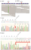
Genetic analysis of index patient (III-4). (A,B) Integrated genome view of DSG2-p.S363X and TBX20-p.D278X, respectively. (C,D) Electropherograms of DSG2-p.S363X and TBX20-p.D278X.
Figure 6
Figure 6
Cell transfection experiments reveal absence of truncated DSG2 at the plasma membrane. HT-1080 cells were transfected with pEYFP–N1–DSG2 (WT = wild-type) or the truncated pEYFP–N1–DSG2–p.S363X plasmids. The fluorescence signal of DSG2-EYFP is shown in yellow. N-Cadherin was costained using primary and Cy3-conjugated secondary antibodies and is shown in red. DAPI was used for staining of the nuclei and is shown in blue. Scale bars represent 10 µm. Of note, the truncated form of DSG2 is not localized at the plasma membrane.

References

    1. Stöllberger C., Wegner C., Finsterer J. Left ventricular hypertrabeculation/noncompaction, cardiac phenotype, and neuromuscular disorders. Herz. 2019;44:659–665. doi: 10.1007/s00059-018-4695-1. - DOI - PubMed
    1. Myasnikov R.P., Kulikova O.V., Meshkov A.N., Kiseleva A.V., Shumarina A.O., Koretskiy S.N., Zharikova A.A., Divashuk M.G., Kharlap M.S., Serduk S.E., et al. New variant of MYH7 gene nucleotide sequence in familial non-compaction cardiomyopathy with benign course. Ration. Pharmacother. Cardiol. 2020;16:383–391. doi: 10.20996/1819-6446-2020-06-01. - DOI
    1. Myasnikov R.P., Blagova O.V., Kulikova O.V., Mershina E.A., Kharlap M.S., Andreenko E.Y., Koretsky S.N., Serdyuk S.E., Bazaeva E.V., Virabova I.A., et al. The specifics of noncompacted cardiomyopathy manifestation. Cardiovasc. Ther. Prev. 2015;14:78. doi: 10.15829/1728-8800-2015-5-78-82. - DOI
    1. Li S., Zhang C., Liu N., Bai H., Hou C., Wang J., Song L., Pu J. Genotype-positive status is associated with poor prognoses in patients with left ventricular noncompaction cardiomyopathy. J. Am. Heart Assoc. 2018;7 doi: 10.1161/JAHA.118.009910. - DOI - PMC - PubMed
    1. Kulikova O.V., Myasnikov R.P., Mershina E.A., Pilus P.S., Koretskiy S.N., Meshkov A.N., Kiseleva A.V., Kharlap M.S., Sinitsyn V.E., Sdvigova N.A., et al. The familial form of non-compaction cardiomyopathy: Types of myocardial remodeling, clinical course. Multicenter register results. Ther. Arch. 2021;94 doi: 10.26442/00403660.2021.04.200677. - DOI

Publication types