. 2021 Jun 24;11(7):1661.
doi: 10.3390/nano11071661.

Combining Augmented Radiotherapy and Immunotherapy through a Nano-Gold and Bacterial Outer-Membrane Vesicle Complex for the Treatment of Glioblastoma

Affiliations

Combining Augmented Radiotherapy and Immunotherapy through a Nano-Gold and Bacterial Outer-Membrane Vesicle Complex for the Treatment of Glioblastoma

Mei-Hsiu Chen et al. Nanomaterials (Basel). .

Abstract

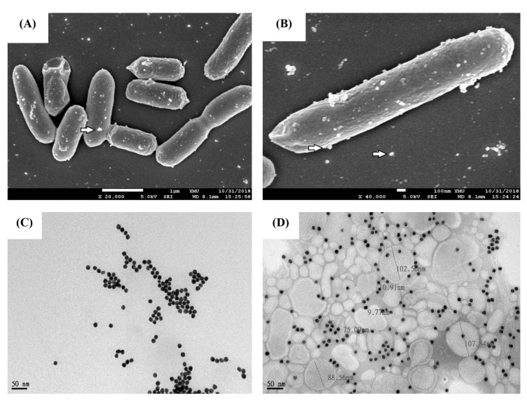
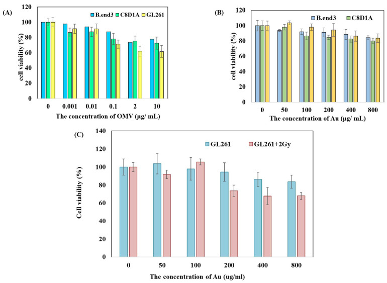
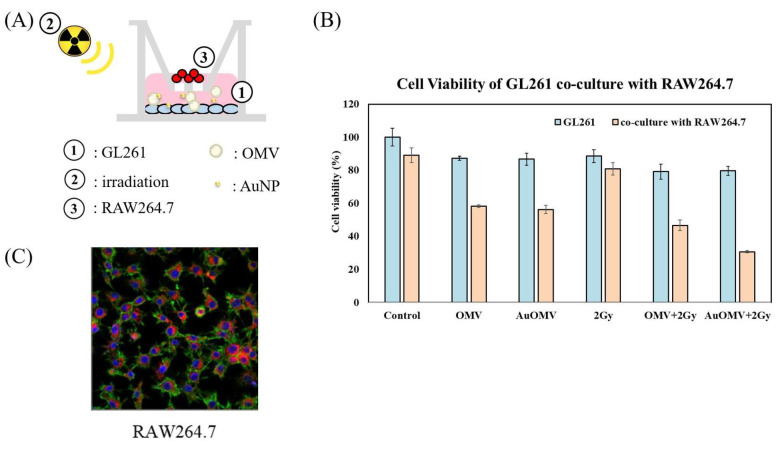
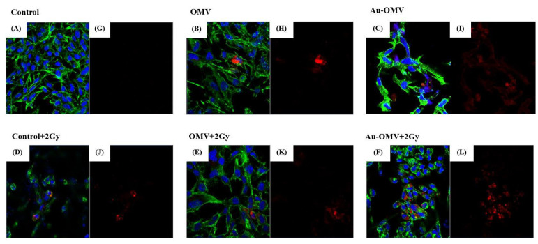
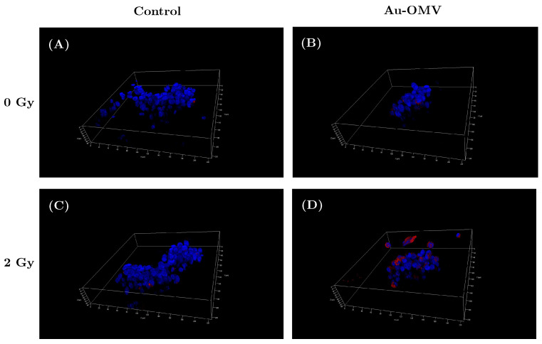
Glioblastoma, formerly known as glioblastoma multiforme (GBM), is refractory to existing adjuvant chemotherapy and radiotherapy. We successfully synthesized a complex, Au-OMV, with two specific nanoparticles: gold nanoparticles (AuNPs) and outer-membrane vesicles (OMVs) from E. coli. Au-OMV, when combined with radiotherapy, produced radiosensitizing and immuno-modulatory effects that successfully suppressed tumor growth in both subcutaneous G261 tumor-bearing and in situ (brain) tumor-bearing C57BL/6 mice. Longer survival was also noted with in situ tumor-bearing mice treated with Au-OMV and radiotherapy. The mechanisms for the successful treatment were evaluated. Intracellular reactive oxygen species (ROS) greatly increased in response to Au-OMV in combination with radiotherapy in G261 glioma cells. Furthermore, with a co-culture of G261 glioma cells and RAW 264.7 macrophages, we found that GL261 cell viability was related to chemotaxis of macrophages and TNF-α production.

Keywords: glioblastoma; gold nanoparticles; immunotherapy; outer membrane vesicles; radioenhancer.


Conflict of interest statement

The authors declare that they have no known competing financial interests or personal relationships that could have appeared to influence the work reported in this paper. The funders had no role in the design of the study; in the collection, analyses, or interpretation of data; in the writing of the manuscript, or in the decision to publish the results.

Figures

Figure 1
Figure 1
Characterizations of Au–OMV nanoparticles. (A,B) Scanning electron microscope image (SEM) of OMV production by E. coli. (C,D) TEM images of Au and Au–OMV.
Figure 2
Figure 2
Toxicity of (A) OMV and (B) Au to B.end3 mouse brain endothelial cells, C8D1A mouse astrocytes, and GL261 mouse glioma cells. (C) Au augmented the radiotoxic effect towards GL261 mouse glioma cells.
Figure 3
Figure 3
(A) GL261 glioma cells (in lower well) were treated with various treatments and co-cultured with RAW 264.7 Macrophages (in upper well) using 0.4 μm pored transwells. (B) GL261 cell viability in response to various treatments when co-cultured for 24 h with RAW 264.7 cells. (C) Co-culture in transwell system with a 3 μm porous membrane at the bottom of the upper chamber. Migrations of RAW 264.7 mouse macrophages towards glioma cells (chemoatxis of microphages) were noted and were labeled with VybrantTM DiD (red) in the lower chamber.
Figure 4
Figure 4
Confocal microscopy of immunofluorescence imaging of the GL 261 glioma cells co-cultured with RAW 264.7 macrophages. (A,G) Control and treatments with (B,H) OMV, (C,I) Au–OMV, (D,J) 2 Gy X-ray, (E,K) OMV + 2 Gy X-ray, and (F,L) Au–OMV + 2 Gy X-ray. ( immunofluorescences: H33342/DAPI (blue for nucleus), phalloidin (green for actin), and VybrantTM DiD (red for macrophages).
Figure 5
Figure 5
Intracellular ROS production of G261 glioma cells in response to (A) different concentrations of OMV and (B) various treatments.
Figure 6
Figure 6
ROS production of live G261 glioma cells grown on 3D spheres in control (A), in response to (B) Au–OMV, (C) 2-Gy radiotherapy, and (D) the combination of Au–OMV and radiotherapy under confocal microscopy. The live cells were labeled with H33342/DAPI (blue for nucleus), phalloidin (green for actin), and CellROXTM Deep Red Reagent for oxidative stress detection (pink for ROS).
Figure 7
Figure 7
Western blot of TNF-α released from RAW 264.7 macrophages co-cultured with GL261 glioma cells. GAPDH was used as a loading control.
Figure 8
Figure 8
(A) Schematic diagram of subcutaneous OMV or Au–OMV injections with radiation therapy for GL261 glioma tumor-bearing mice. (B,C) Tumor volume kept growing in the control group, but the growth was suppressed in all treatment groups, especially in the group treated with Au–OMV + 2 Gy radiotherapy, whose tumors did not grow, even at the end of the experiment.
Figure 9
Figure 9
(A) Schematic diagram of intracranial Au–OMV injection with radiation therapy for GL261 glioma tumor-bearing mice. (B) Assessment of the IVIS imaging system as a method for monitoring tumor growth in a GL261 in situ tumor mode. IVIS imaging of mouse brain tumors on days 5, 12, 15, and 18 after implantation of tumor cells; imaging results of three mice in each group. All images were at the same scale. (C) Kaplan–Meier survival curves of control and treatment groups (N > 5 per arm).

References

    1. Nørøxe D.S., Poulsen H.S., Lassen U. Hallmarks of glioblastoma: A systematic review. ESMO Open. 2016;1:e000144. doi: 10.1136/esmoopen-2016-000144. - DOI - PMC - PubMed
    1. Mujokoro B., Adabi M., Sadroddiny E., Adabi M., Khosravani M. Nano-structures mediated co-delivery of therapeutic agents for glioblastoma treatment: A review. Mater. Sci. Eng. 2016;69:1092–1102. doi: 10.1016/j.msec.2016.07.080. - DOI - PubMed
    1. Šamec N., Zottel A., Videtič Paska A., Jovčevska I. Nanomedicine and Immunotherapy: A Step Further towards Precision Medicine for Glioblastoma. Molecules. 2020;25:490. doi: 10.3390/molecules25030490. - DOI - PMC - PubMed
    1. Schuemann J., Bagley A.F., Berbeco R., Bromma K., Butterworth K.T., Byrne H.L., Chithrani B.D., Cho S.H., Cook J.R., Favaudon V., et al. Roadmap for metal nanoparticles in radiation therapy: Current status, translational challenges, and future directions. Phys. Med. Biol. 2020;65:21RM02. doi: 10.1088/1361-6560/ab9159. - DOI - PubMed
    1. Haume K., Rosa S., Grellet S., Śmiałek M.A., Butterworth K.T., Solov’yov A.V., Prise K.M., Golding J., Mason N.J. Gold nanoparticles for cancer radiotherapy: A review. Cancer Nanotechnol. 2016;7:8. doi: 10.1186/s12645-016-0021-x. - DOI - PMC - PubMed

LinkOut - more resources