. 2021 Jun 24;22(13):6780.
doi: 10.3390/ijms22136780.

Perfusable System Using Porous Collagen Gel Scaffold Actively Provides Fresh Culture Media to a Cultured 3D Tissue

Affiliations

Perfusable System Using Porous Collagen Gel Scaffold Actively Provides Fresh Culture Media to a Cultured 3D Tissue

Chikahiro Imashiro et al. Int J Mol Sci. .

Abstract

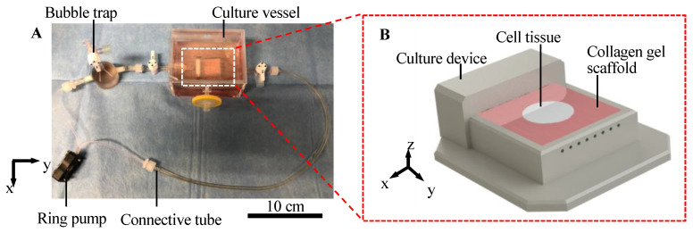
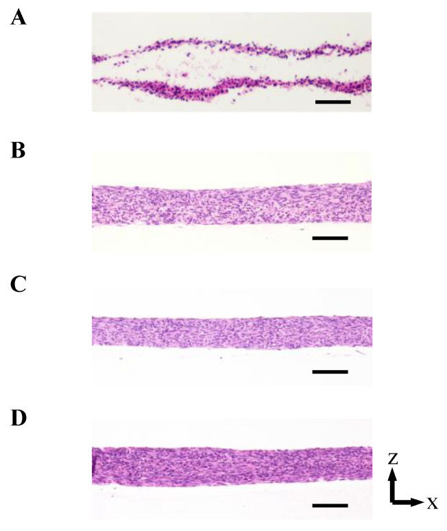
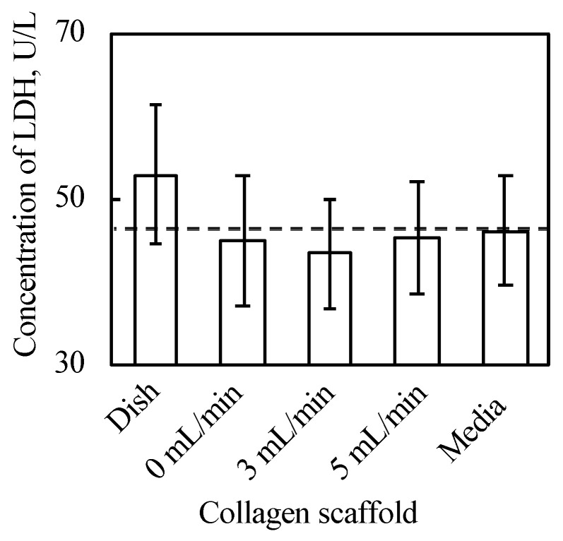
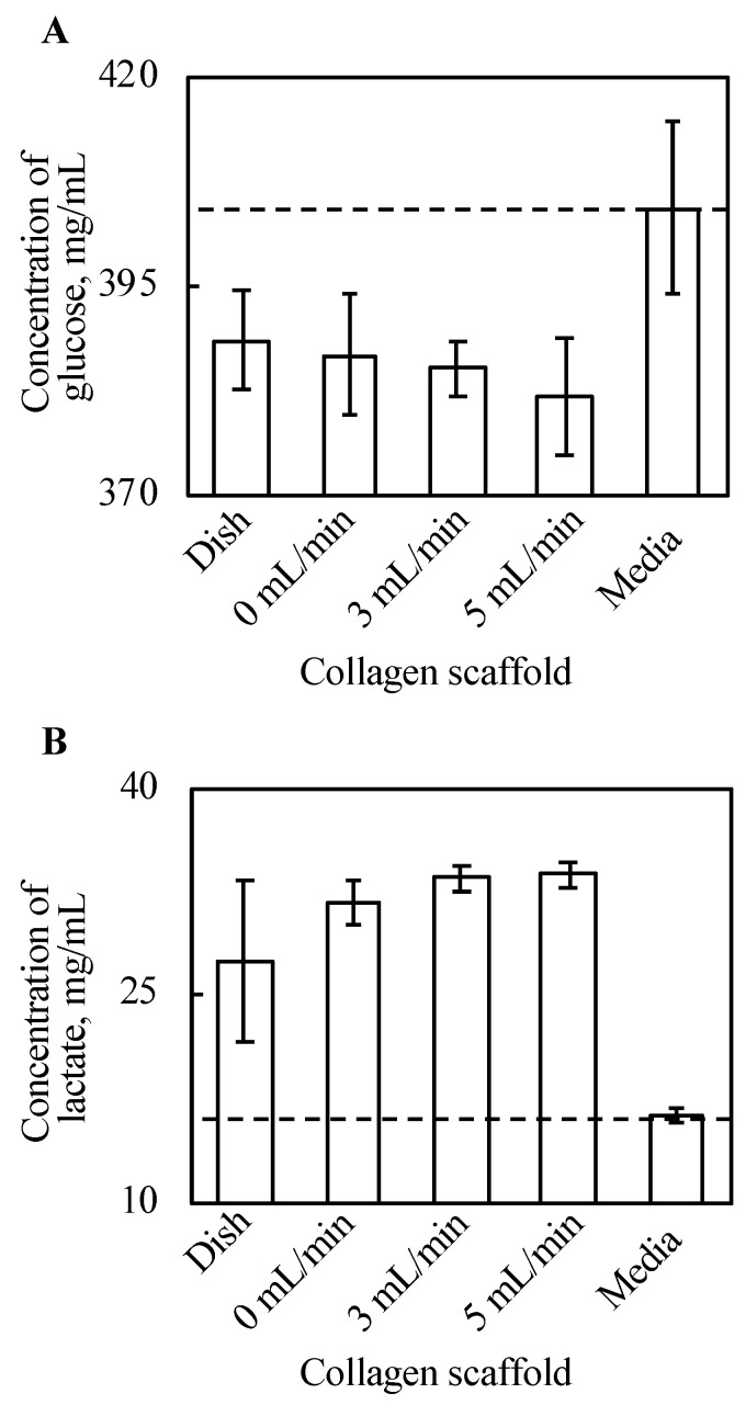
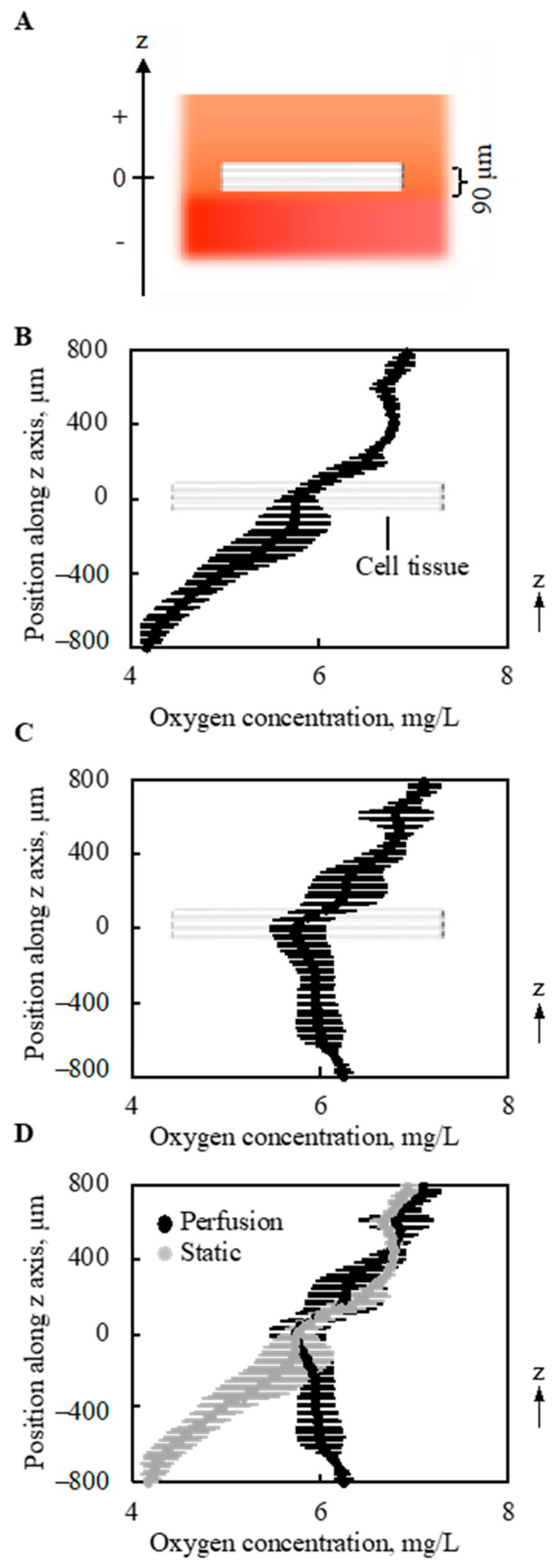
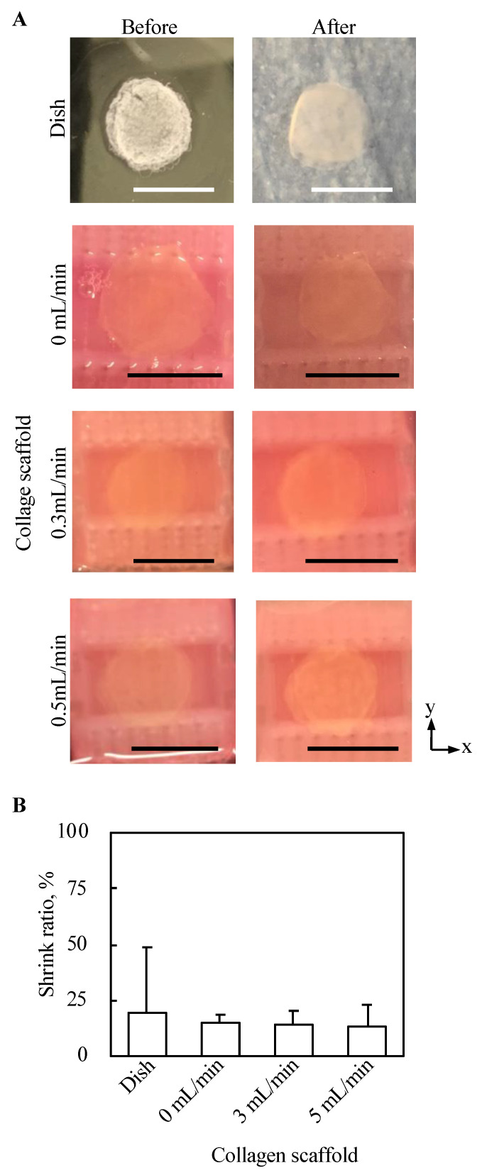
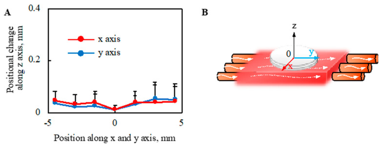
Culturing three-dimensional (3D) tissues with an appropriate microenvironment is a critical and fundamental technology in broad areas of cutting-edge bioengineering research. In addition, many technologies have engineered tissue functions. However, an effective system for transporting nutrients, waste, or oxygen to affect the functions of cell tissues has not been reported. In this study, we introduce a novel system that employs diffusion and convection to enhance transportation. To demonstrate the concept of the proposed system, three layers of normal human dermal fibroblast cell sheets are used as a model tissue, which is cultured on a general dish or porous collagen scaffold with perfusable channels for three days with and without the perfusion of culture media in the scaffold. The results show that the viability of the cell tissue was improved by the developed system. Furthermore, glucose consumption, lactate production, and oxygen transport to the tissues were increased, which might improve the viability of tissues. However, mechanical stress in the proposed system did not cause damage or unintentional functional changes in the cultured tissue. We believe that the introduced culturing system potentially suggests a novel standard for 3D cell cultures.

Keywords: 3D tissue culture; cell sheet technology; convection-diffusion equation; perfusion system.


Conflict of interest statement

T.S. was a shareholder of CellSeed, Inc., Tokyo, Japan. Tokyo Women’s Medical University receives research funds from CellSeed, Inc. Tokyo Women’s Medical University and Waseda University receives research funds from CKD Corporation.

Figures

Figure A1
Figure A1
Procedure showing the preparation of the collagen scaffold.
Figure A2
Figure A2
The densities of the cell tissue cultured for three days. HE-stained cross-section views with the cell tissue culture on a general dish (A) and collagen gel with a 0.5-mL/min perfusion rate (B). Images of 1 and 2 were taken from different samples, and the sample is as shown in Figure 3. Scale bars indicate 100 µm.
Figure A3
Figure A3
The schematic image shows the evaluation methods for shear stress from the media flow (left upper side) and the tensile deformation due to the leaky flow toward the bottom of the tissue.
Figure 1
Figure 1
The concept of the perfusion system. Comparison of conventional culture (A) and the ideal culture (B) environment. Schematic images of the conventional culture method (C) and the culture method realized in this study (D).
Figure 2
Figure 2
Illustration of the culture system. Overview of the whole perfusion system (A). Schematic image of culture device with the collagen scaffold (B).
Figure 3
Figure 3
The densities of the cell tissue cultured for 3 d with each method. HE stained cross-section views with the cell tissue culture on a general dish (A) and collagen gel with perfusion rates of 0 (B), 0.3 (C), and 0.5 (D) mL/min. Scale bars indicate 100 µm. Figure A2 in Appendix B shows the reproducibility of the results with the cell tissue culture on a general dish and collagen gel with a 0.5-mL/min perfusion rate.
Figure 4
Figure 4
Results of LDH assay (n = 3~7, mean ± SD). The cell tissue cultured on ubiquitous culture dish released LDH in the media. However, cell tissues cultured using the other methods did not show the release.
Figure 5
Figure 5
The results of glucose assay (A) and lactate assay (B) (n = 3~7, mean ± SD). The y-axis represents the concentration of each substance in the supernatant with each culture method and incubated DMEM without cells. The cell tissue cultured on a collagen gel scaffold shows a higher glucose consumption and lactate production. Further, along with increasing perfusion speed, both the glucose consumption and lactate production increased.
Figure 6
Figure 6
Oxygen concentration around and in the tissue (n = 3). The schematic image showing measured region (A). Without perfusion, a gradual decrease in the oxygen concentration was shown along with the depth direction (B). However, owing to the perfusion, the minimal point of oxygen concentration was at the location of the cell tissue (C). On the other hand, the density of oxygen itself around the cell tissue is very similar. The results shown in (B,C) were overlapped for comparison (D). Note that the perfusion speed is 5 mL/min.
Figure 7
Figure 7
Evaluation of the morphology from the top view of tissue cultured using each method. The macro top-view images of the tissues (A). The shrink ratios of tissues are shown (n = 3~7, mean ± SD) (B).
Figure 8
Figure 8
Evaluation of morphological change in the direction of the z axis owing to the perfusion. The positional change of the cell tissue surface owing to perfusion (A) is shown (n = 3) (details are shown in Figure A3). Note that the schematic image to show each axis is shown (B). Note that the perfusion speed was 5 mL/min in this figure.

References

    1. Sekine H., Okano T. Capillary Networks for Bio-Artificial Three-Dimensional Tissues Fabricated Using Cell Sheet Based Tissue Engineering. Int. J. Mol. Sci. 2021;22:92. doi: 10.3390/ijms22010092. - DOI - PMC - PubMed
    1. Sarkiri M., Fox S.C., Fratila-Apachitei L.E., Zadpoor A.A. Bioengineered Skin Intended for Skin Disease Modeling. Int. J. Mol. Sci. 2019;20:1407. doi: 10.3390/ijms20061407. - DOI - PMC - PubMed
    1. Furuhashi M., Morimoto Y., Shima A., Nakamura F., Ishikawa H., Takeuchi S. Formation of contractile 3D bovine muscle tissue for construction of millimetre-thick cultured steak. NPJ Sci. Food. 2021;5:1–8. doi: 10.1038/s41538-021-00090-7. - DOI - PMC - PubMed
    1. Post M.J. Cultured meat from stem cells: Challenges and prospects. Meat Sci. 2012;92:297–301. doi: 10.1016/j.meatsci.2012.04.008. - DOI - PubMed
    1. Wang X.-Y., Jin Z.-H., Gan B.-W., Lv S.-W., Xie M., Huang W.-H. Engineering interconnected 3D vascular networks in hydrogels using molded sodium alginate lattice as the sacrificial template. Lab Chip. 2014;14:2709–2716. doi: 10.1039/C4LC00069B. - DOI - PubMed

LinkOut - more resources