Review
. 2021 Jun 20;13(6):1178.
doi: 10.3390/v13061178.

The Viral Capsid: A Master Key to Access the Host Nucleus

Affiliations
Review

The Viral Capsid: A Master Key to Access the Host Nucleus

Guillermo Blanco-Rodriguez et al. Viruses. .

Abstract

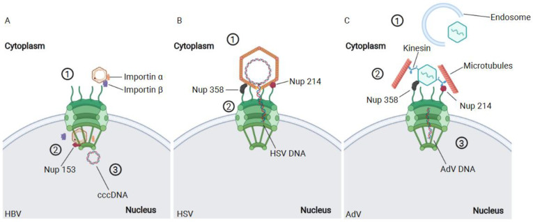
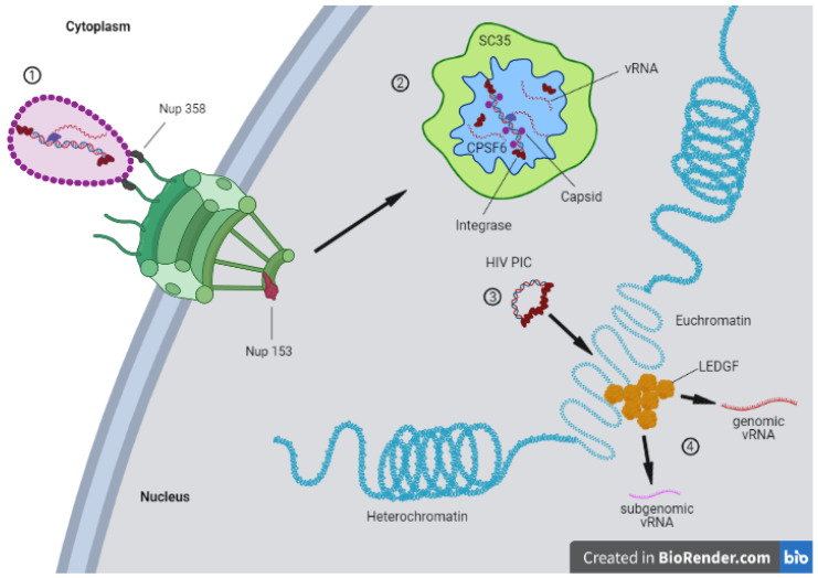
Viruses are pathogens that have evolved to hijack the cellular machinery to replicate themselves and spread to new cells. During the course of evolution, viruses developed different strategies to overcome the cellular defenses and create new progeny. Among them, some RNA and many DNA viruses require access to the nucleus to replicate their genome. In non-dividing cells, viruses can only access the nucleus through the nuclear pore complex (NPC). Therefore, viruses have developed strategies to usurp the nuclear transport machinery and gain access to the nucleus. The majority of these viruses use the capsid to manipulate the nuclear import machinery. However, the particular tactics employed by each virus to reach the host chromatin compartment are very different. Nevertheless, they all require some degree of capsid remodeling. Recent notions on the interplay between the viral capsid and cellular factors shine new light on the quest for the nuclear entry step and for the fate of these viruses. In this review, we describe the main components and function of nuclear transport machinery. Next, we discuss selected examples of RNA and DNA viruses (HBV, HSV, adenovirus, and HIV) that remodel their capsid as part of their strategies to access the nucleus and to replicate.

Keywords: host–viral interactions; viral nuclear entry; viral replication.


Conflict of interest statement

The authors declare no conflict of interest.

Figures

Figure 1
Figure 1
Different viruses employ various strategies in order to access to the nucleus. (A) The HBV travels to the vicinity of the NPC protected by its capsid. Thus, it employs importin α and β to access the nucleus (1). The small size of HBV capsid permits the import of this structure complete to the nucleus. The uncoating of HBV capsid takes place assisted by Nup 153 (2) releasing the genome in the nucleus of the cell (3). (B) The HSV capsid is too large to be imported complete to the nucleus. Therefore, the HSV binds to the cytoplasmic filaments of NPC via Nup358 and Nup214 (1). Once the capsid docks at the NPC, the viral DNA is injected to the nucleus (2). (C) The adenovirus uncoating begins during the release of the viral capsid from the endosomes (1) and is completed at the NPC (2), where there is a complex formed by the capsid, Nup 358, Nup214, microtubules, and kinesin. The mechanical stress caused by the binding to microtubules and to the NPC contributes to completing the tear down of adenovirus capsids. Eventually, the viral DNA is released in the nucleus (3). Created with BioRender.com, accessed on 7 June 2021.
Figure 2
Figure 2
The inhibition of HIV-1 retrotranscription by nevirapine does not block nuclear import of HIV. (A) The use of nevirapine (10 µM) efficiently inhibits the reverse transcription and therefore the replication of HIV-1. This blockage does not impede the import of HIV-1 integrase to the nucleus at 6 h post-infection, as the fluorescence microscopy image shows (antibody against the IN-HA tagged has been employed with a secondary antibody conjugated with Alexa 647). (B) Transmission electron microscopy (TEM) coupled to immuno-gold labeling (antibodies anti CA and anti IN-HA, respectively recognized by secondary antibodies conjugated with gold particles of 10 nm and 5 nm) reveals the presence of HIV-1 integrase and capsid complexes in the nucleus of infected cells in the presence or in absence of nevirapine. Black arrows represent capsid, whereas integrase is pointed out by the white arrows. p-values with <0.0001 are represented by **** than Images adapted from https://www.pasteur.fr/fr/journal-recherche/actualites/technologie-hiv-1-anchor-devoile-vih-1-cellules-vivantes-lors-etape-entree-nucleaire, accessed on 2 June 2021and results corroborating the Blanco-Rodriguez et al. study [106].
Figure 3
Figure 3
The HIV-1 core carrying the viral genome docks at the NPC followed by nuclear translocation (1). The nuclear RT takes place in HIV-1 MLOs formed by nuclear speckles factors and CPSF6 (2). These are maturation sites of the pre-integration complex (PIC), which once formed is released from them to join euchromatin regions containing LEDGF clusters (3). These are proviral sites where the viral genome is transcribed in genomic and subgenomic viral RNAs (4). Created with BioRender.com, accessed on 7 June 2021.

References

    1. Cronshaw J.M., Krutchinsky A.N., Zhang W., Chait B.T., Matunis M.J. Proteomic analysis of the mammalian nuclear pore complex. J. Cell Biol. 2002;158:915–927. doi: 10.1083/jcb.200206106. - DOI - PMC - PubMed
    1. Gall J.G. Octagonal nuclear pores. J. Cell Biol. 1967;32:391–399. doi: 10.1083/jcb.32.2.391. - DOI - PMC - PubMed
    1. Löschberger A., Linde S., van de Dabauvalle M.-C., Rieger B., Heilemann M., Krohne G., Sauer M. Super-resolution imaging visualizes the eightfold symmetry of gp210 proteins around the nuclear pore complex and resolves the central channel with nanometer resolution. J. Cell Sci. 2012;125:570–575. doi: 10.1242/jcs.098822. - DOI - PubMed
    1. Lučić V., Förster F., Baumeister W. Structural studies by electron tomography: From cells to molecules. Annu. Rev. Biochem. 2005;74:833–865. doi: 10.1146/annurev.biochem.73.011303.074112. - DOI - PubMed
    1. Wan W., Briggs J. Cryo-electron tomography and subtomogram averaging. Methods Enzymol. 2016;579:329–367. doi: 10.1016/bs.mie.2016.04.014. - DOI - PubMed

Publication types

LinkOut - more resources