Review
. 2021 Jun 28;22(13):6924.
doi: 10.3390/ijms22136924.

The Roles of Pseudophosphatases in Disease

Affiliations
Review

The Roles of Pseudophosphatases in Disease

Andrew M Mattei et al. Int J Mol Sci. .

Abstract

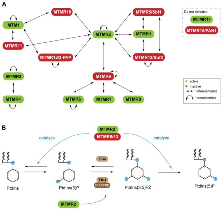
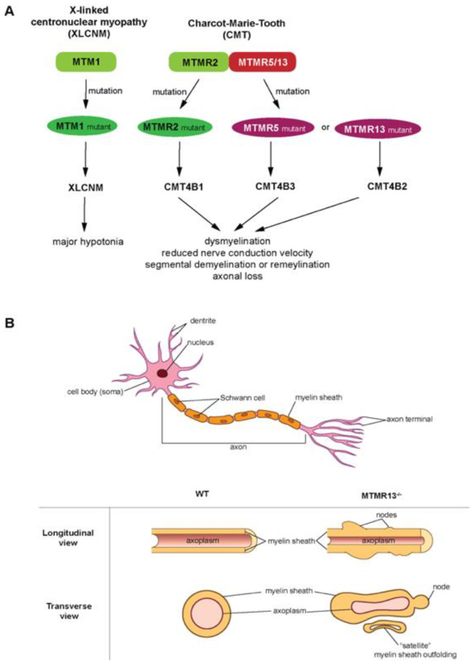
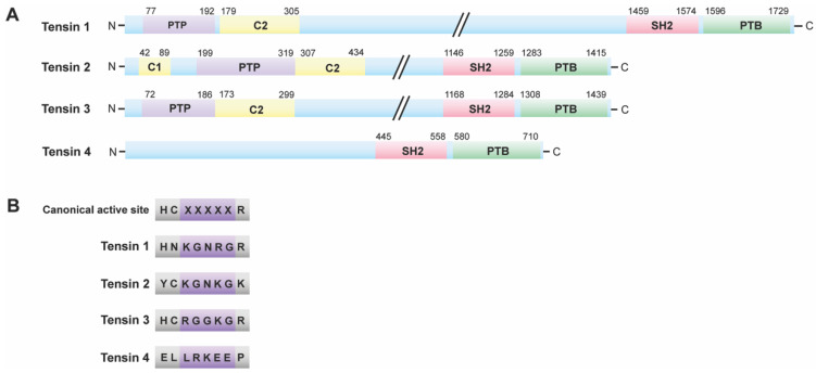
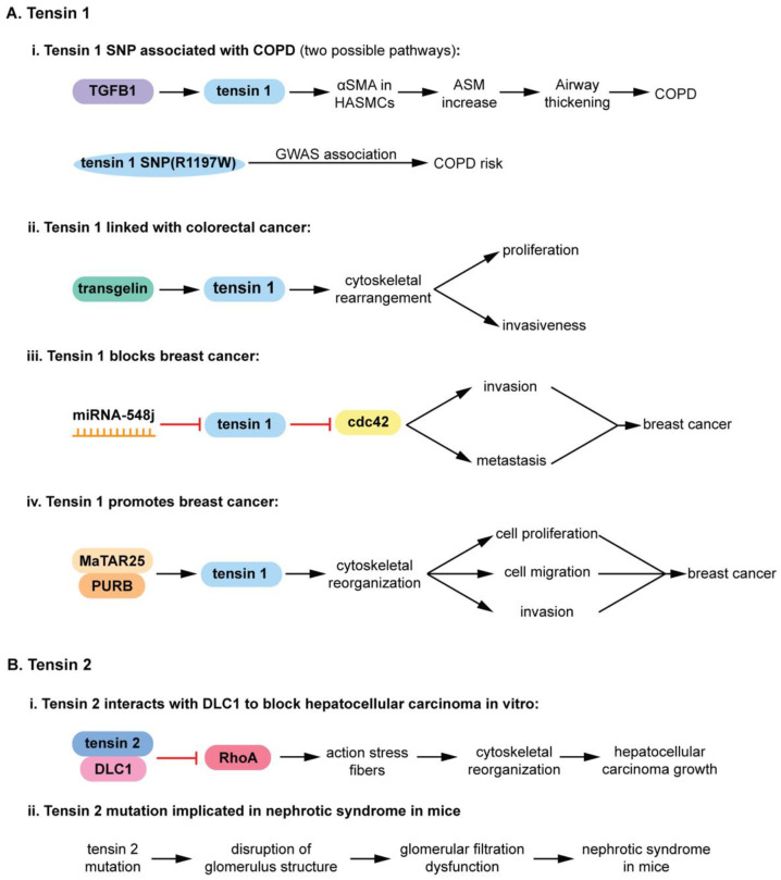
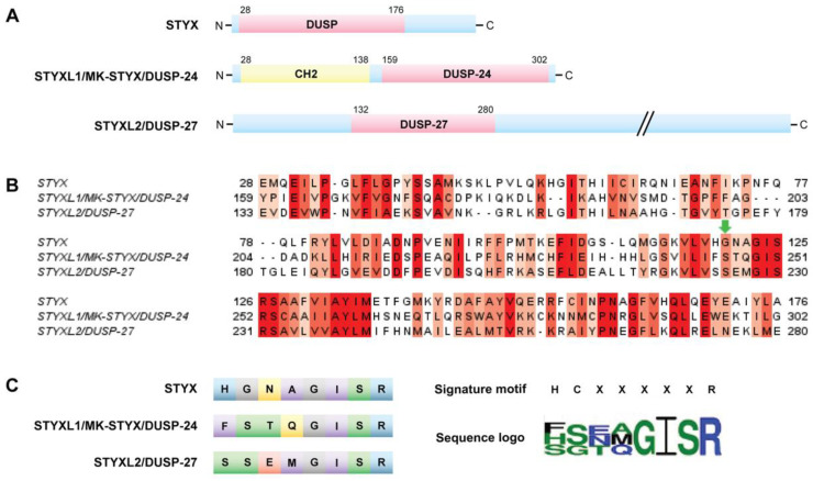
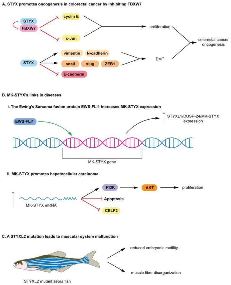
The pseudophosphatases, atypical members of the protein tyrosine phosphatase family, have emerged as bona fide signaling regulators within the past two decades. Their roles as regulators have led to a renaissance of the pseudophosphatase and pseudoenyme fields, catapulting interest from a mere curiosity to intriguing and relevant proteins to investigate. Pseudophosphatases make up approximately fourteen percent of the phosphatase family, and are conserved throughout evolution. Pseudophosphatases, along with pseudokinases, are important players in physiology and pathophysiology. These atypical members of the protein tyrosine phosphatase and protein tyrosine kinase superfamily, respectively, are rendered catalytically inactive through mutations within their catalytic active signature motif and/or other important domains required for catalysis. This new interest in the pursuit of the relevant functions of these proteins has resulted in an elucidation of their roles in signaling cascades and diseases. There is a rapid accumulation of knowledge of diseases linked to their dysregulation, such as neuropathies and various cancers. This review analyzes the involvement of pseudophosphatases in diseases, highlighting the function of various role(s) of pseudophosphatases involvement in pathologies, and thus providing a platform to strongly consider them as key therapeutic drug targets.

Keywords: MK-STYX (MAPK (mitogen-activated protein kinase) phosphoserine/threonine/tyrosine-binding protein); STYX (phosphoserine/threonine/tyrosine-interacting protein); disease; dual specificity phosphatases (DUSPs); myotubularin phosphatases (MTMs); protein tyrosine phosphatases (PTPs); pseudoenzymes; pseudophosphatases; tensin.


Conflict of interest statement

The authors declare no conflict of interests.

Figures

Figure 1
Figure 1
Dimerization states of the myotubularin (MTM) family: (A) Active and catalytically inactive forms are indicated in green and red, respectively. Active and inactive coupling (heterodimerization) is common; however, self-association (homodimerization) has also been reported among both active and inactive MTMs. MTMR14 and MTMR15 are not known to dimerize. (B) Phosphorylation schema for phosphatidylinositol (PtdIns), a molecule important for endosomal-lysosomal membrane trafficking, with example PtdIns kinases (PIKFYVE) and non-MTM phosphatases (FIG4), along with active and inactive MTMs (labelled green and red, respectively). Heterodimerization of an active (MTMR2) and inactive (MTMR5, MTMR13) leads to a stabilized complex with PtdIns phosphatase potential. MTMR2 and FIG4 interactions have been implicated in vivo, though further characterization is necessary.
Figure 2
Figure 2
Myotubularins (MTMs) in diseases: (A) X-linked centronuclear myopathy (XLCNM) and Charcot-Marie-Tooth (CMT), pathologies associated with MTM family mutations. XLCNM is a rare congenital myopathy characterized by decreased muscle tone or weakness (hypotonia), and is caused by mutations to the MTM1 gene. CMT denotes a group of peripheral nerve neuropathies with an incidence of ~1 in 2500 people, and is characterized by abnormal myelin folding, diminished nerve conduction velocities, as well as peripheral axon loss. CMT type 4B (CMT4B) is caused by mutations in the MTM family proteins (MTMR2, MTMR5 or MTMR13) which lead to CMT4B1, CMT4B3 or CMT4B2, respectively. (B) (Top) Canonical representation of a PNS neuron and its various processes. (Bottom) Idealized illustration adopted from Robinson et al. [30] of the aberrant myelin phenotypes associated with CMT4B2 (longitudinal and transverse views of MTMR13-/- knockout axons) as compared to Wild Type (WT) axons. Abnormal myelination, as depicted by “satellite” myelin outfoldings, as well as myelin “nodes”, are present in mouse models deficient of MTMR13, resulting in diminished nerve conduction velocities typical of CMT4B patients.
Figure 3
Figure 3
Tensin family: (A) Structure of tensin family members. All tensin family members have a C-terminal PTB (phosphotyrosine-binding) domain preceded by an SH2 (Src 2Homology 2) domain. These structures vary beyond the SH2 and PTB domains. (B) Active sites of tensin family members. Some tensin family members have active site motifs that differ from the catalytically active canonical PTP HCX5R motif. Tensin 1 and 2 are the pseudophosphatase members of the family, whereas tensin 3 possibly retains catalytic competence. Tensin 4 lacks a PTP domain; therefore, it is neither a phosphatase nor a pseudophosphatase.
Figure 4
Figure 4
Roles of tensin family members in disease: (A) Tensin 1 roles in disease. (i) Tensin 1 SNP is linked to chronic obstructive pulmonary disease (COPD) thorough experimental evidence and GWAS. TGFβ1 upregulates tensin 1 (R1197W) expression, increasing downstream effectors leading to airway thickening that is characteristic of COPD. (ii) Tensin 1 is linked with human colorectal cancer through transgelin signaling, upregulating metastatic and proliferative signaling of colorectal cancer cells. (iii) Tensin 1. Tensin 1 translation is prevented by miRNA-548j, resulting in hyperactivation of cdc42 that increases invasion and metastasis in breast cancer. (iv) Tensin 1 promotes breast cancer. The MaTAR25 lncRNA interacts with PURB to increase tensin 1 mRNA and protein levels, resulting in cytoskeletal rearrangements that augment proliferation, migration, and invasion in breast cancer cells. (B) Tensin 2 roles in disease. (i) Tensin 2-DLC1 (deleted in liver cancer 1) complex inhibits RhoA. This prevents RhoA-mediated actin stress fiber generation, which in turn prevents cytoskeletal rearrangements that support hepatocellular carcinoma growth. (ii) Tensin 2 mutation implicated in nephrotic syndrome in mice. An 8-nucleotide deletion in tensin 2 triggers a frame shift that introduces a nonsense mutation. This mutant gene is associated with lower tensin 2 mRNA and protein levels in mouse kidneys. These tensin 2 deficient mice present with disruption of glomerulus structure and glomerular filtration dysfunction, suggesting that tensin 2 deficiency leads to nephrotic syndrome in an animal disease model.
Figure 5
Figure 5
STYX pseudophosphatases: (A) Structure of STYX pseudophosphatases. STYX has an N-terminal DUSP domain. MK-STYX features an N-terminal CH2 domain and a C-terminal DUSP domain. STYXL2 contains an N-terminal DUSP domain. (B) Sequence alignment of the phosphatase domains of the STYX pseudophosphatases (Clustal Omega 1.2.4). Conserved residues are shown in red, where darker red indicates higher conservation (Jalview 2.11.14 with a threshold of >5.5). The green arrow indicates where the essential active-site cysteine residue would be located in a catalytically active PTP (HCX5R). (C) The left-hand side of the panel compares the active site sequences of the STYX pseudophosphatases. Amino acid color indicates chemical property. Polar amino acids are shown in green, neutral in yellow, basic in blue, acidic in red, hydrophobic in purple, and glycine in grey. The right-hand side of the panel shows the sequence of the consensus active signature motif of PTPs compared to the sequence logo of the three STYX pseudophosphatases. The sequence logo was built by WebLogo 3.7.4 with a 2.0-bit scale.
Figure 6
Figure 6
Roles of STYX domain pseudophosphatases in disease: (A) Prototypical STYX roles in colorectal cancer. STYX promotes oncogenesis in colorectal cancer by inhibiting FBXW7. STYX binds FBXW7, preventing other interactions of FBXW7, which is a substrate recruiter for a ubiquitin protein ligase complex. Thus, STYX prevents the degradation of cyclin E and c-Jun, promoting proliferation in colorectal cancer. STYX overexpression increases the expression of vimentin, N-cadherin, snail, slug, and ZEB1, but reduction in E-cadherin. These proteins support EMT; STYX may promote the oncogenesis of colorectal cancer by positively regulating EMT. (B) MK-STYX’s links in diseases. (i) The Ewing’s sarcoma fusion protein EWS-FLI1 increases MK-STYX expression. The EWS-FLI1 oncoprotein binds an ETS binding motif within the MK-STYX gene and increases the MK-STYX’s expression. (ii) MK-STYX promotes hepatocellular carcinoma. An increase in MK-STYX mRNA expression leads to upregulation of PI3K/AKT pathway proteins and an enhancement of proliferation in hepatocellular carcinoma, while inhibiting apoptosis and CELF2. (C) STYXL2 mutation leads to muscular system malfunction. A transgene integration into the STYXL2 gene reduces STYXL2 expression in zebrafish, resulting in reduced embryonic motility, low spontaneous coiling movements, and severely reduced touch response, as well as major muscle fibers disorganization.

References

    1. Murphy J.M., Farhan H., Eyers P.A. Bio-Zombie: The rise of pseudoenzymes in biology. Biochem. Soc. Trans. 2017;45:537–544. doi: 10.1042/BST20160400. - DOI - PubMed
    1. Todd A.E., Orengo C.A., Thornton J.M. Sequence and structural differences between enzyme and nonenzyme homologs. Structure. 2002;10:1435–1451. doi: 10.1016/S0969-2126(02)00861-4. - DOI - PubMed
    1. Murphy J.M., Mace P.D., Eyers P.A. Live and let die: Insights into pseudoenzyme mechanisms from structure. Curr. Opin. Struct. Biol. 2017;47:95–104. doi: 10.1016/j.sbi.2017.07.004. - DOI - PubMed
    1. Hinton S.D. The role of pseudophosphatases as signaling regulators. Biochim. Biophys. Acta Mol. Cell Res. 2019;1866:167–174. doi: 10.1016/j.bbamcr.2018.07.021. - DOI - PubMed
    1. Reiterer V., Pawlowski K., Desrochers G., Pause A., Sharpe H.J., Farhan H. The dead phosphatases society: A review of the emerging roles of pseudophosphatases. FEBS J. 2020;287:4198–4220. doi: 10.1111/febs.15431. - DOI - PubMed

MeSH terms

LinkOut - more resources