Review
. 2021 Jun 28;22(13):6937.
doi: 10.3390/ijms22136937.

Emerging Advances of Nanotechnology in Drug and Vaccine Delivery against Viral Associated Respiratory Infectious Diseases (VARID)

Affiliations
Review

Emerging Advances of Nanotechnology in Drug and Vaccine Delivery against Viral Associated Respiratory Infectious Diseases (VARID)

Amir Seyfoori et al. Int J Mol Sci. .

Abstract

Viral-associated respiratory infectious diseases are one of the most prominent subsets of respiratory failures, known as viral respiratory infections (VRI). VRIs are proceeded by an infection caused by viruses infecting the respiratory system. For the past 100 years, viral associated respiratory epidemics have been the most common cause of infectious disease worldwide. Due to several drawbacks of the current anti-viral treatments, such as drug resistance generation and non-targeting of viral proteins, the development of novel nanotherapeutic or nano-vaccine strategies can be considered essential. Due to their specific physical and biological properties, nanoparticles hold promising opportunities for both anti-viral treatments and vaccines against viral infections. Besides the specific physiological properties of the respiratory system, there is a significant demand for utilizing nano-designs in the production of vaccines or antiviral agents for airway-localized administration. SARS-CoV-2, as an immediate example of respiratory viruses, is an enveloped, positive-sense, single-stranded RNA virus belonging to the coronaviridae family. COVID-19 can lead to acute respiratory distress syndrome, similarly to other members of the coronaviridae. Hence, reviewing the current and past emerging nanotechnology-based medications on similar respiratory viral diseases can identify pathways towards generating novel SARS-CoV-2 nanotherapeutics and/or nano-vaccines.

Keywords: COVID-19; SARS-CoV-2; nano-vaccine; nanomedicine; respiratory disease; viral infection.


Conflict of interest statement

The authors declare no conflict of interest.

Figures

Figure 1
Figure 1
(A) Nasal virus entry homing in the nasopharynx cavity and virus attachment on epithelial cells and olfactory neurons. Virus replication in olfactory cells can decrease the ability of smell sensing and cause inflammation in the nasopharynx. (B) Oral cavity, salivary component including dimeric IgA, cathepsins, and sublingual and laryngeal lymph nodes are the first line of lymphoid tissue and antibody production. (C) Oral-nasal virus entry, oropharynx cavity, and virus attachment on the epithelial cells of throat. (D) Normal alveoli in first days of virus entry: thin layer of fibroblasts, low density and distribution of immune cells in a single epithelial layer, eosinophil and neutrophils number in a normal range. (E) Severe infection in the alveolar region: macrophages became foam cells. Inflammatory agents induce mucus secretion and increase the viscosity of the mucosal barrier. Alveolar epithelial cells die via apoptosis or viral cytolysis, NK cells increment, and neutrophils induce a cytokine storm. (F) Inflammatory conditions induce fibrosis and fibroblast cells proliferation, which can cause thickness of the alveolar cavity, resulting in respiratory distress. (G) Lung obstruction results in decreased respiratory rate.
Figure 2
Figure 2
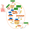
Proposed immune escape mechanism of SARS-CoV, MERS-CoV and possibly SARS-CoV-2. SARS-CoV-2 is attached to its receptor on the surface of target ACE2 positive cells, such as alveolar or other target cells, reducing the anti-viral IFN responses, leading to viral replication and propagation. COVID-19 may inhibit the pathways induced by TLRs3, 7, and 8, which are expressed in the endosomes. The suppression of these molecules leads to dampening of NF-kB, IRF signaling cascades, and STAT1/2 function in the nucleus, which decreases in the production of Type I IFNs responses. Delayed Type I IFNs responses may trigger immune exhaustion and the invasion of neutrophils and monocytes/macrophages into the infected cell, which may lead to cytokine storms and Th2 type responses resulting in poor outcomes.
Figure 3
Figure 3
Mechanisms of vaccine administration using nanoparticles in VARID. (A) Intranasal vaccination: The aerosol-based nanoparticles containing the mRNA of virus antigen is transferred through the mucus layer into the nasal epithelial tissues by micro-fold cells (M cells) or passively through epithelial cell junctions. Nanoparticles are captured by DCs, and alveolar macrophages (AMQ) are passed by epithelial junctions and by other APCs, such as B cells. The mRNA of the antigen is translated into a specific peptide and presented to immature T cells, activating them and B cells. The activated B cells proliferate in the B cell zone to maturity and enter the systemic circulation to reach the inflammation site. IgA and B cells locally differentiate into antibody-secreting plasma cells to produce IgA dimers. The IgA dimers are secreted via polymeric Ig receptor (pIgR) at the mucosal surface. NALT/BALT immune response induces long-lasting B and T memory cells able to activate a rapid memory response [110]. (B) Other types of nano-vaccine injection, such as intramuscular, subcutaneous, and intravenous, can induce systemic reactions and IgG production, thus inducing lung protection. (C) Some specific nanoparticles induce the immunomodulatory responses using CPN, which can induce the IgG and specific CTL production against antigens. Systemic injection of nanoparticles can induce iBALT and local responses.

References

    1. Sohrabi C., Alsafi Z., O’Neill N., Khan M., Kerwan A., Al-Jabir A., Iosifidis C., Agha R. World Health Organization declares global emergency: A review of the 2019 novel coronavirus (COVID-19) Int. J. Surg. 2020;76:71–76. doi: 10.1016/j.ijsu.2020.02.034. - DOI - PMC - PubMed
    1. van Doorn H.R., Yu H. Viral Respiratory Infections. Hunt. Trop. Med. Emerg. Infect. Dis. 2020:284–288. doi: 10.1016/B978-0-323-55512-8.00033-8. - DOI
    1. Ibricevic A., Pekosz A., Walter M.J., Newby C., Battaile J.T., Brown E.G., Holtzman M.J., Brody S.L. Influenza virus receptor specificity and cell tropism in mouse and human airway epithelial cells. J. Virol. 2006;80:7469–7480. doi: 10.1128/JVI.02677-05. - DOI - PMC - PubMed
    1. Sureda A., Alizadeh J., Nabavi S.F., Berindan-Neagoe I., Cismaru C.A., Jeandet P., Los M.J., Clementi E., Nabavi S.M., Ghavami S. Endoplasmic reticulum as a potential therapeutic target for covid-19 infection management? Eur. J. Pharmacol. 2020;882:173288. doi: 10.1016/j.ejphar.2020.173288. - DOI - PMC - PubMed
    1. Zhang H., Penninger J.M., Li Y., Zhong N., Slutsky A.S. Angiotensin-converting enzyme 2 (ACE2) as a SARS-CoV-2 receptor: Molecular mechanisms and potential therapeutic target. Intensive Care Med. 2020;46:586–590. doi: 10.1007/s00134-020-05985-9. - DOI - PMC - PubMed

MeSH terms