Review
. 2021 Jun 28;11(7):478.
doi: 10.3390/membranes11070478.

On the Coupling between Mechanical Properties and Electrostatics in Biological Membranes

Affiliations
Review

On the Coupling between Mechanical Properties and Electrostatics in Biological Membranes

Vanesa Viviana Galassi et al. Membranes (Basel). .

Abstract

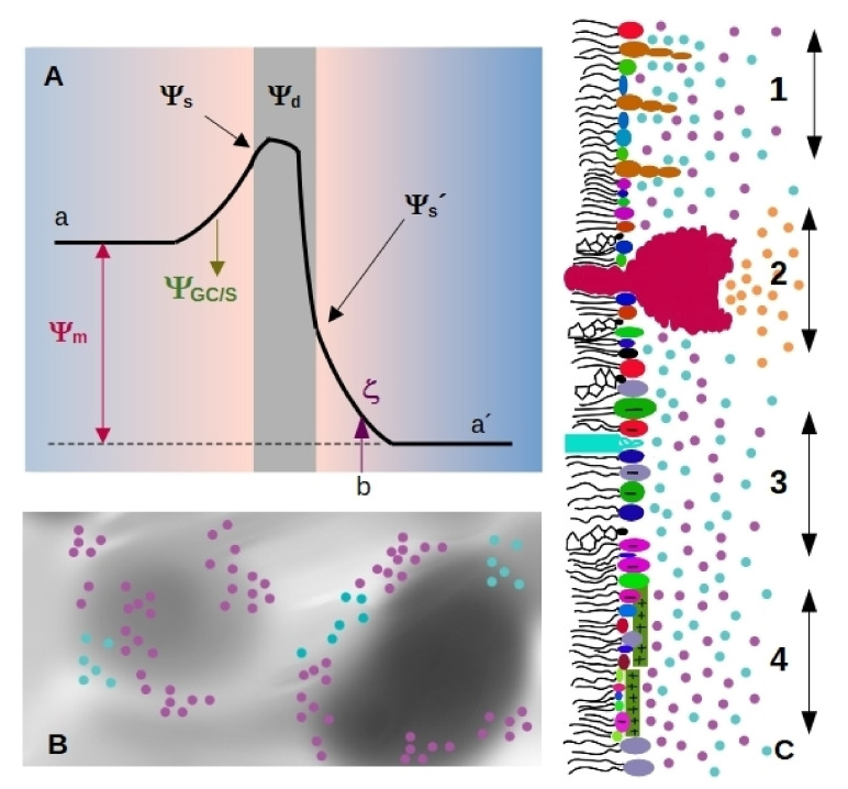
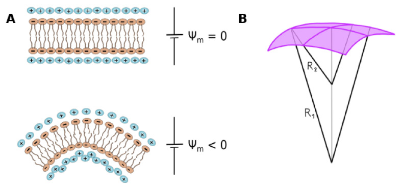
Cell membrane structure is proposed as a lipid matrix with embedded proteins, and thus, their emerging mechanical and electrostatic properties are commanded by lipid behavior and their interconnection with the included and absorbed proteins, cytoskeleton, extracellular matrix and ionic media. Structures formed by lipids are soft, dynamic and viscoelastic, and their properties depend on the lipid composition and on the general conditions, such as temperature, pH, ionic strength and electrostatic potentials. The dielectric constant of the apolar region of the lipid bilayer contrasts with that of the polar region, which also differs from the aqueous milieu, and these changes happen in the nanometer scale. Besides, an important percentage of the lipids are anionic, and the rest are dipoles or higher multipoles, and the polar regions are highly hydrated, with these water molecules forming an active part of the membrane. Therefore, electric fields (both, internal and external) affects membrane thickness, density, tension and curvature, and conversely, mechanical deformations modify membrane electrostatics. As a consequence, interfacial electrostatics appears as a highly important parameter, affecting the membrane properties in general and mechanical features in particular. In this review we focus on the electromechanical behavior of lipid and cell membranes, the physicochemical origin and the biological implications, with emphasis in signal propagation in nerve cells.

Keywords: electric field; electro-mechanical properties; electroporation; flexoelectricity; lipid ionization; nerve impulse.


Conflict of interest statement

The authors declare no conflict of interest.

Figures

Figure 1
Figure 1
Scheme illustrating membrane electrostatics. (A) Electrostatic potential profiles that bear a membrane (gray region) along the normal axis. Volta potentials are usually different on each side of the membrane, the difference between the value on the left (a) and right (a’) side corresponds to the membrane potential Ψm. Charged species on the membrane generate the surface potential Ψs. The charged membrane induce an ion cloud and a potential drop characterized through Gouy-Chapman or Stern model ΨGC/S. In the scheme, the slipping plane is at a distance b from the membrane, and the potential value at this point corresponds to the zeta potential ζ. Membranes are composed of multipoles organized in an ordered array, generating the dipole potential Ψd. (B) Possible charge distribution in the membrane plane with non-homogeneous electrical properties. Gray levels indicate different values for the dielectric constant. Cyan circles represent cations and pink circles, anions. (C) Lateral view of a membrane hemilayer, where different situations coexist. As in B, cyan circles represent cations and pink circles, anions. 1—Gangliosides protrude from the plane formed by the polar head groups of less bulky lipids, generating a rough surface. 2—A pump or a channel generates a local ion gradient (orange circles correspond to protons, sodium, calcium or the specific ion that passes trough the pump/channel). 3—Region of the membrane enriched in anionic lipids. This region will attract cations. 4—cationic peptides adsorbed to a region of the membrane generate a positive surface that attracts anions.
Figure 2
Figure 2
Peptide-membrane interaction. (A) Interaction of cationic peptides with lipid monolayers composed of fatty acids. The incorporation (measured as an increase in lateral pressure Δπ) depends on the monolayer dipole potential (ΔΦ0), and is maximal for systems with inverted (negative) values. Measurements were performed using perfluorotetradecanoic acid (PFTD), myristic acid (MA) or palmitic acid (PA). The plot is adapted with permission from [78]. Copyright (2018) American Chemical Society. (B) Shape fluctuations of GUVs in the presence of increasing concentration of a cationic peptide. Adapted from [82]. (C) Kinetic of membrane nanotube retraction formed from a GUV by means of optical tweezers. The effect of peptide addition is shown at different times. Adapted from [84] with permision from Elsevier. Copyright Elsevier (2021).
Figure 3
Figure 3
Flexoelectricity. (A) Scheme of flexoelectricity: Membrane is plane in the absence of a membrane electric field (Ψm=0), while a curvature is induced due to the effect of the non-zero potential. (B) Scheme of curvature definition in a bidimensional plane, where R1 and R2 are the two main curvature radii.
Figure 4
Figure 4
Scheme showing the relation between membrane phase state and excitability. (A) The cell membrane is excitable if the resting state (asterisk) is in the vicinity of an ordered-disordered transition (e.g., from liquid expanded (LE) to liquid condensed phase (LC) in liquid monolayers; or from lamellar fluid to gel in lipid bilayers). This is illustrated by the P-V isotherm and the derived isothermal compressibility on the right (the arrow illustrates the respective slice through the phase plane). State changes move the system state (asterisk) through phase space and hence change the physical properties of the membrane. At low T / high P the membrane “freezes” into a crystalline-like state. (B) In a T-pH plot, the phase boundary is sigmoidal. The underlying reason for this additional nonlinearity is that the headgroups of membrane molecules are ionizable. This results in a nonlinear change of the transition temperature with pH. Thus, acidification at constant T and P can move the resting state into the LC phase. (C) According to the melting point depression theory [162,166], anesthetics leave the resting state in the disordered phase, but increase its distance to the transition. This figure is reproduced from ref. [164] with permission from Elsevier.

Similar articles

Cited by

References

    1. Israelachvili J.N. Intermolecular and Surface Forces. Academic Press; Cambridge, MA, USA: 2011.
    1. Montich G.G., Bustos M.M., Maggio B., Cumar F.A. Micropolarity of interfaces containing anionic and neutral glycosphingolipids as sensed by Merocyanine 540. Chem. Phys. Lipids. 1985;38:319–326. doi: 10.1016/0009-3084(85)90026-X. - DOI
    1. Cooke R., Kuntz I. The properties of water in biological systems. Annu. Rev. Biophys. Bioeng. 1974;3:95–126. doi: 10.1146/annurev.bb.03.060174.000523. - DOI - PubMed
    1. Mild K.H., Løvtrup S. Movement and structure of water in animal cells. Ideas and experiments. Biochim. Et Biophys. Acta Rev. Biomembr. 1985;822:155–167. doi: 10.1016/0304-4157(85)90006-1. - DOI - PubMed
    1. Bagatolli L.A., Stock R.P., Olsen L.F. Coupled response of membrane hydration with oscillating metabolism in live cells: An alternative way to modulate structural aspects of biological membranes? Biomolecules. 2019;9:687. doi: 10.3390/biom9110687. - DOI - PMC - PubMed