. 2021 Jun 28;9(7):744.
doi: 10.3390/biomedicines9070744.

BPC 157 Therapy and the Permanent Occlusion of the Superior Sagittal Sinus in Rat: Vascular Recruitment

Affiliations

BPC 157 Therapy and the Permanent Occlusion of the Superior Sagittal Sinus in Rat: Vascular Recruitment

Slaven Gojkovic et al. Biomedicines. .

Abstract

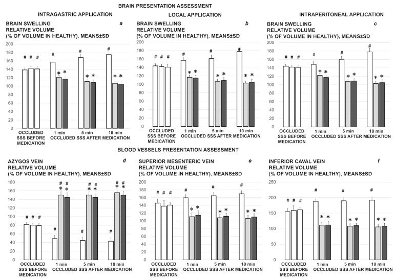
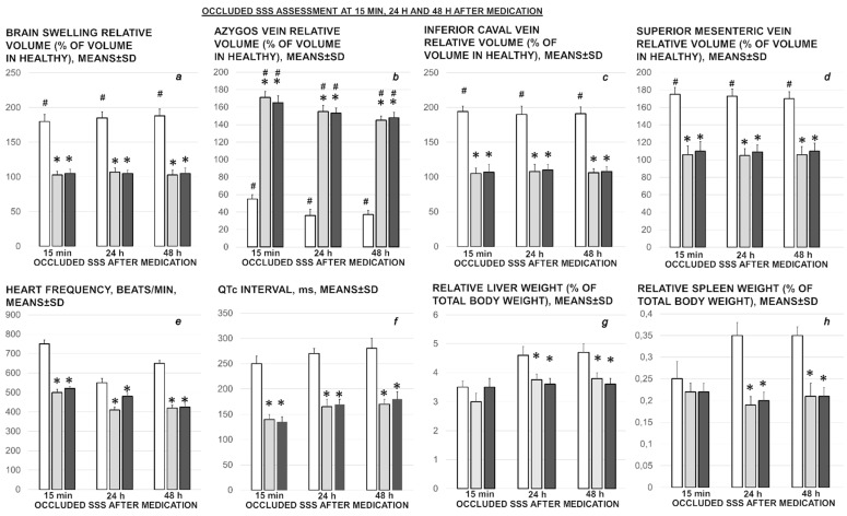
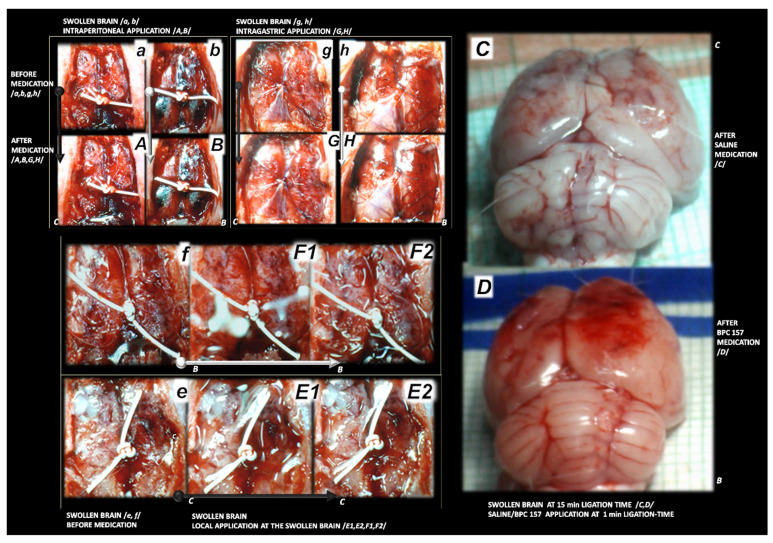
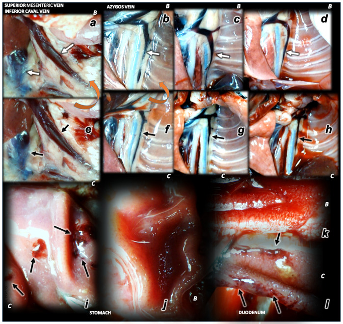
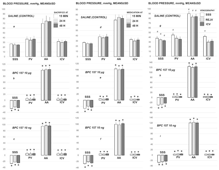
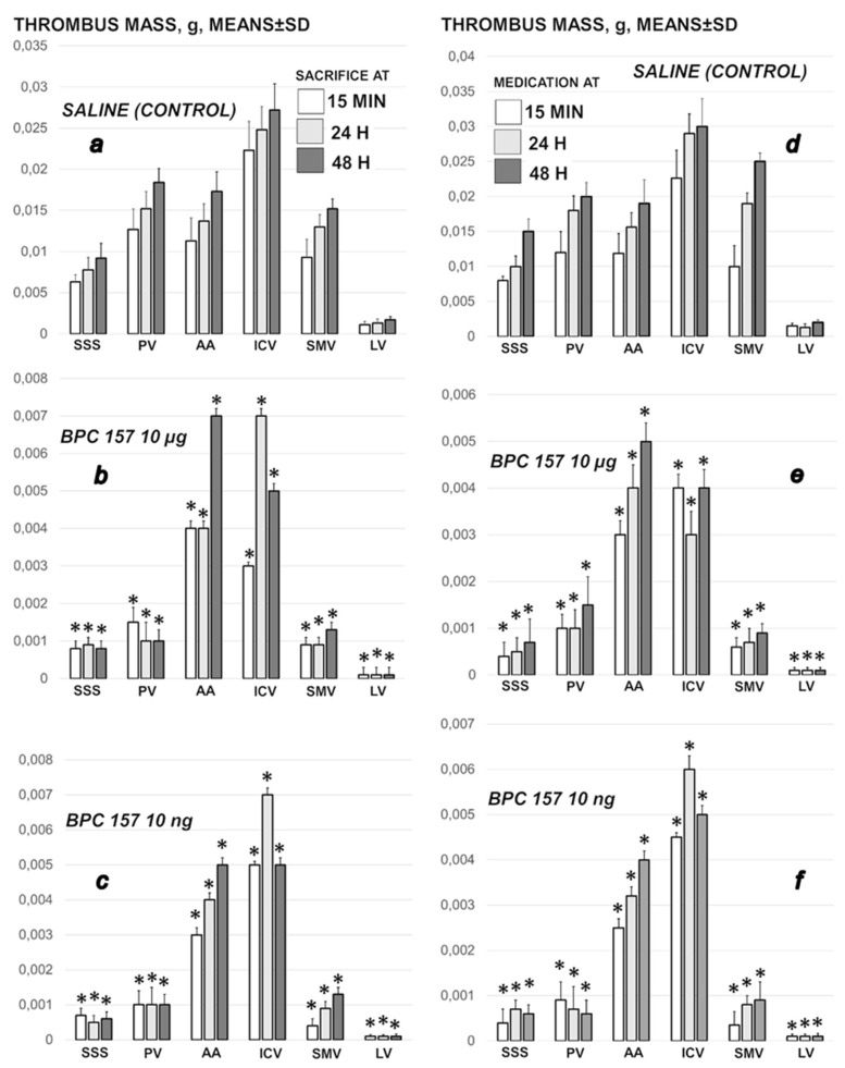
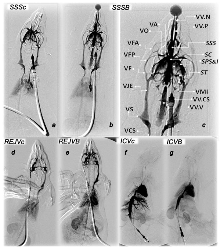
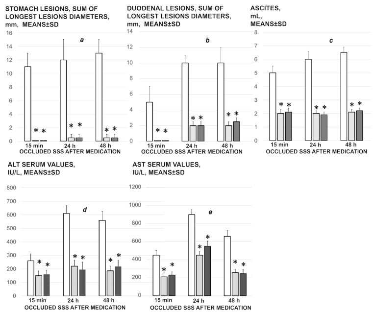
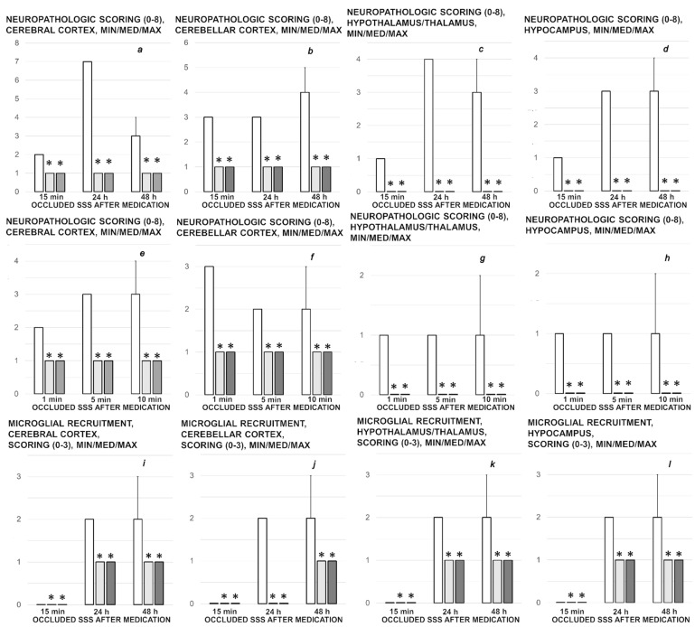
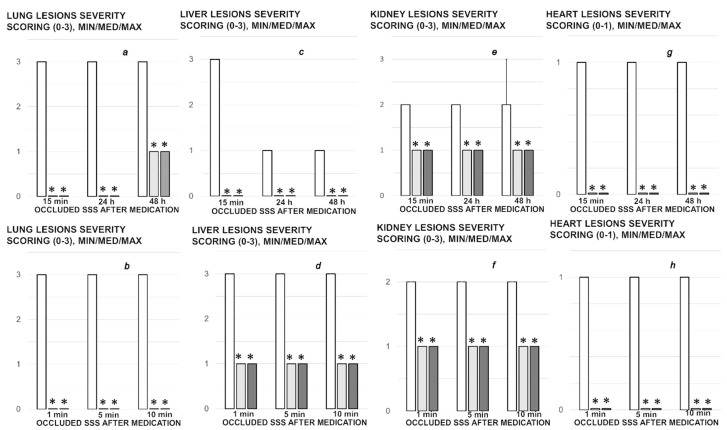
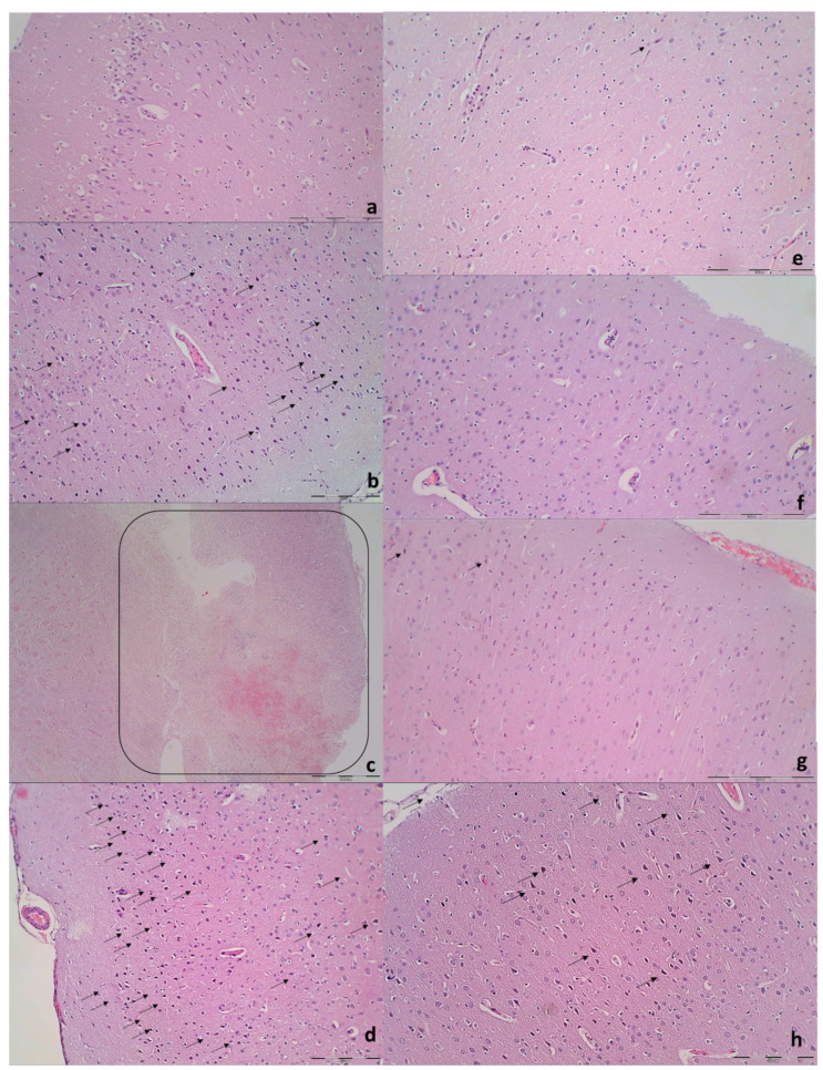
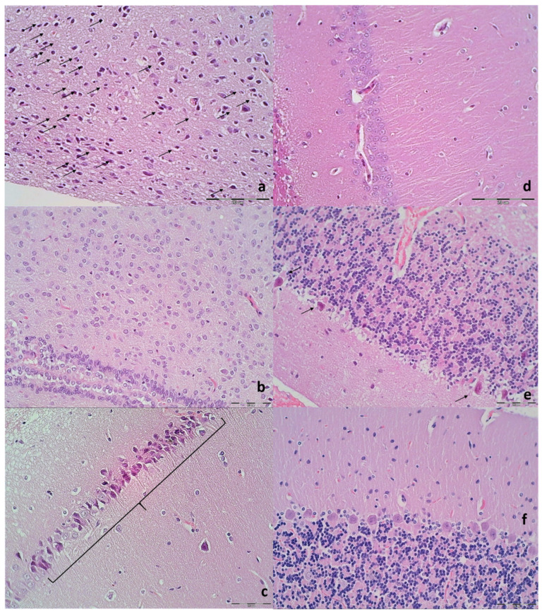
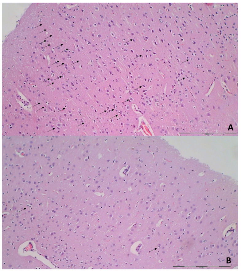
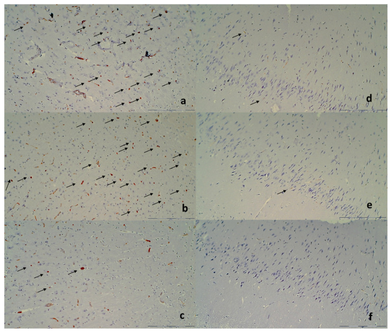
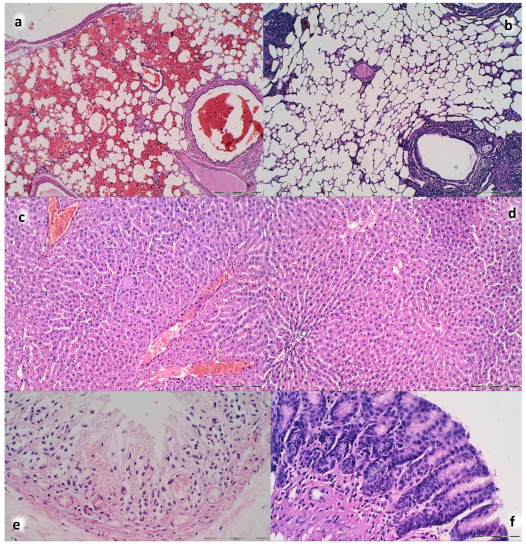
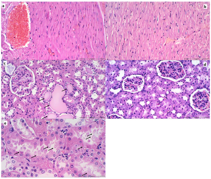
We show the complex syndrome of the occluded superior sagittal sinus, brain swelling and lesions and multiple peripheral organs lesions in rat. Recovery goes centrally and peripherally, with the stable gastric pentadecapeptide BPC 157, which alleviated peripheral vascular occlusion disturbances, rapidly activating alternative bypassing pathways. Assessments were gross recording, venography, ECG, pressure, microscopy, biochemistry. The increased pressure in the superior sagittal sinus, portal and caval hypertension, aortal hypotension, arterial and venous thrombosis, severe brain swelling and lesions (cortex (cerebral, cerebellar), hypothalamus/thalamus, hippocampus), particular veins (azygos, superior mesenteric, inferior caval) dysfunction, heart dysfunction, lung congestion as acute respiratory distress syndrome, kidney disturbances, liver failure, and hemorrhagic lesions in gastrointestinal tract were all assessed. Rats received BPC 157 medication (10 µg/kg, 10 ng/kg) intraperitoneally, intragastrically, or topically to the swollen brain at 1 min ligation-time, or at 15 min, 24 h and 48 h ligation-time. BPC 157 therapy rapidly attenuates the brain swelling, rapidly eliminates the increased pressure in the ligated superior sagittal sinus and the severe portal and caval hypertension and aortal hypotension, and rapidly recruits collateral vessels, centrally ((para)sagittal venous collateral circulation) and peripherally (left superior caval vein azygos vein-inferior caval vein). In conclusion, as shown by all assessments, BPC 157 acts against the permanent occlusion of the superior sagittal sinus and syndrome (i.e., brain, heart, lung, liver, kidney, gastrointestinal lesions, thrombosis), given at 1 min, 15 min, 24 h or 48 h ligation-time. BPC 157 therapy rapidly overwhelms the permanent occlusion of the superior sagittal sinus in rat.

Keywords: BPC 157; occlusion; rats; superior sagittal sinus; therapy; vascular recruitment.


Conflict of interest statement

The authors declare that there are no conflict of interest.

Figures

Figure 1
Figure 1
Brain and blood vessels presentation assessed as the relative volume, % of volume in healthy, before ligation (100%), and then in the rats with occluded superior sagittal sinus, immediately after ligation (occluded superior sagittal sinus (SSS) before medication, white bars), and then after therapy (occluded superior sagittal sinus (SSS) after medication) at 1 min, 5 min, and 10 min ligation-time. Therapy was saline 5 mL/kg (white bars), BPC 157 10 µg/kg (light gray bars), 10 ng/kg (dark gray bars). In the assessment of the brain swelling progress or elimination, the medication effect was shown when given intragastrically (a), locally at the swollen brain (b), or intraperitoneally (c); or intragastrically with azygos (d), superior mesenteric (e), and inferior caval vein (f) assessment (while the effects of the intraperitoneal and local application regimens, providing the similar results were not specifically shown). Means ± SD, * p ˂ 0.05, at least vs. corresponding control; # p ˂ 0.05, at least vs. regular healthy values.
Figure 2
Figure 2
Brain (a) and blood vessels (azygos vein (b), inferior caval vein (c), superior mesenteric vein (d)) presentation assessed as the relative volume, % of volume in healthy, before ligation (100%), and then in the rats with occluded superior sagittal sinus (SSS), after therapy at 15 min, 24 h, and 48 h ligation-time. Heart frequency, beats/min (e), and QTc interval, ms (f), relative liver (g) and spleen (h) weight (% of total body weight) (low, right) at 15 min, 24 h, and 48 h ligation-time. Therapy was saline 5 mL/kg (white bars), BPC 157 10 µg/kg (light gray bars), 10 ng/kg (dark gray bars) given intragastrically at 1 min ligation time while the effects of the intraperitoneal and local application regimens, providing the similar results were not specifically shown. Means ± SD, * p ˂ 0.05, at least vs. corresponding control; # p ˂ 0.05, at least vs. regular healthy values (100%).
Figure 3
Figure 3
Rat brain swelling presentation at 15 min ligation time in rats with superior sagittal sinus ligation. Rats received saline (C) or BPC 157 (B) and indicated with small white C and small white B close to the corresponding presentation. Swollen brain before medication (a,b,eh), and after medication, given earlier at 1 min ligation-time (right, (C,D)). Therapy applied at 15 min ligation-time (A,B,E1,E2,F1,F2,G,H), presented the effect of medication at 5 min therapy-time (medication was given intraperitoneally (A,B) (upper, left, saline (small (C)), BPC 157 (small (B)), intragastrically (G,H) (upper, middle, saline (small (C)), BPC 157 (small (B)). Local application at the swollen brain. Swollen brain before medication (e,f). For medication given locally at the swollen brain (arrows), 30 s therapy-time (brain under solution, small C (control, saline), small B (BPC 157 solution)) and 45 s therapy-time (immediately upon solution resorption, small C (control, saline), small B (BPC 157 solution)) (low, (E1,E2) (saline), (F1,F2) (BPC 157)). Aggravation of the swelling, saline (intraperitoneally) (A), intragastrically (G); saline (topically) (E1) (30 s therapy-time), (E2) immediately upon solution resorption (45 s therapy-time). Attenuated swelling, BPC 157 (intraperitoneally (B); intragastrically (H) (at 5 min therapy-time); BPC 157 (topically) (F1) (30 s therapy-time), (F2) immediately upon solution resorption (45 s therapy-time). (C,D). Characteristic gross brain presentation with medication saline (C) or BPC 157 (D) given intraperitoneally at 1 min ligation time.
Figure 4
Figure 4
Illustrative presentation of blood vessels (superior mesenteric vein, inferior caval vein, azygos vein) (ah), stomach and duodenal lesions (il). Rats received saline (C) or BPC 157 (B). Superior mesenteric vein, inferior caval vein (a,e), azygos vein (bd,f,g). Upon superior sagittal sinus ligation, as illustration of the severe portal and caval hypertension, and not functioning connective pathways, congested superior mesenteric and inferior caval vein (arrows) (e), collapsed azygos vein (arrows) (f). Azygos vein remains weakly presented at 24 h (azygos vein, low, middle, (g)) and 48 h ligation time (azygos vein, low, right, (h)). Upon BPC 157 intragastric application, superior mesenteric and inferior caval veins are rapidly decongested (a) and azygos vein reopen (arrows) (upper, left, (b)) sustainably presented also at 24 h (azygos vein, upper, middle (c)) and 48 h ligation time (azygos vein, upper, right (d)) (the effect along with elimination of the portal/caval hypertension, and elimination of the intracranial hypertension). Low. At 15 min ligation time, hemorrhagic lesions in the duodenum (right (l)) and stomach (left (j)) of the control rats (arrows), and spared mucosa in the duodenum (k) and stomach (i) of the BPC 157 treated rats. Magnification ×30.
Figure 5
Figure 5
Blood pressure of the rats with ligated superior sagittal sinus in the superior sagittal sinus (SSS), portal vein (PV), abdominal aorta (AA) and inferior caval vein (ICV), at the end of the 5 min assessment following medication (BPC 157 10 µg/kg, 10 ng/kg; saline 5 mL/kg) given intraperitoneally. Early medication at 1 min ligation time, blood pressure assessment before euthanized at 15 min (a), 24 h (b) and 48 h (c) ligation time. Delayed medication at 15 min (d), 24 h (e) and 48 h (f) ligation time. (gi) For verification of the effect of venography, assessment at 5 min upon the additional 1 mL of saline application in the superior sagittal sinus (SSS), right external jugular vein (REJV) or inferior caval vein (ICV), inducing venous hypertension additionally originated within the cranium, inside the sinus lumen, neck or abdomen, at 15 min, and subsequent immediate ip application of saline or BPC 157. Similar effects were obtained with BPC 157 intragastric or local application at the swollen brain (data not specifically shown). Means ± SD, * p ˂ 0.05, at least vs. control; # p ˂ 0.05, at least vs. regular saline-control in non-venography studies.
Figure 6
Figure 6
Thrombus presentation in the rats with ligated superior sagittal sinus in the superior sagittal sinus (SSS), portal vein (PV), abdominal aorta (AA), inferior caval vein (ICV), superior mesenteric vein (SMV) and lienal vein (LV) at the end of the 5 min assessment following medication (BPC 157 10 µg/kg, 10 ng/kg; saline 5 mL/kg) given intraperitoneally. Left: Early medication at 1 min ligation time, blood pressure assessment before sacrifice at 15 min, 24 h and 48 h ligation time (ac). Right: Delayed medication at 15 min, 24 h and 48 h ligation time (df). Similar effects were obtained with BPC 157 intragastric or local application at the swollen brain (data not specifically shown). Means ± SD, * p ˂ 0.05, at least vs. control.
Figure 7
Figure 7
Along with saline (c) or BPC 167 (B) presentation of venography in superior sagittal sinus (SSS) (ac), right external jugular vein (REJV) (d,e) and inferior caval vein (ICV) (f,g). SSS. Upper Left: Control venography in superior sagittal sinus (SSSc) (a). Retrograde filling of both jugular external veins and both superior caval veins. Pulmonary congestion with heart dilatation and without inflow of contrast media in the heart. Retrograde filling of pale inferior caval vein. Upper Right: BPC 157 venography in superior sagittal sinus (SSSB) (b,c). Retrograde filling of both jugular external veins and both superior caval veins with vertebral veins. There is no sign of pulmonary congestion and heart dilatation and with normal filling of contrast media in the heart. Retrograde filling of wider inferior caval vein with inflow of hepatic veins, both renal veins with parenchymal phase visualization of both kidneys and adrenals. Presentation of the pterigopalatinal veins (VVP) and nasal veins (VV.N) and presentation of the ophthalmic vein (VO), angularis vein (VA), facial anterior and posterior vein (VFA, VFP), and facial vein (VF), or through cerebri superior veins (VV.CS), sinus cavernosus (SC), sinus petrosus superior and inferior (SPS&I), sinus transversus (ST), through jugular external vein (VJE), subclavia vein (VS) through superior caval veins (VCS). Lower Left: REJV. Control venography through right external jugular vein (REJVc) with retrograde filling of left external jugular vein and left superior caval vein (left, REJVc) (d). There is pulmonary congestion, with insufficient heart contrast filling. Pale visualization of aortic arch and ascendent aorta, without supra-aortic branches. Retrograde filling of inferior caval vein without visualization of renal veins with pale parenchymal phase of both kidneys and adrenals. BPC 157 venography through right external jugular vein (REJVB) (e) with retrograde filling of left external jugular vein and left superior caval vein. Partial visualization of azygos vein. There is no sign of pulmonary congestion, with normal heart contrast filling. Significant visualization of aortic arch, with supra-aortic branches-both common carotid arteries. Clear visualization of thoracic and abdominal aorta. Retrograde filling of dilated inferior caval vein with paravertebral venous plexus. Slight visualization of hepatic veins inflow. Visualization of both renal veins with strong parenchymal phase of both kidneys and adrenals. In addition, there is visualization of portomesenteric veins and intestinal branches. Lower Right: ICV. Control venography through inferior caval vein (ICVc) (f) shows huge congestion within prominent hepatic veins without parenchymal liver phase. In addition, there is severe congestion in pulmonar artery ramification, without parenchymal lung phase and dilatation of the heart with filling of contrast media. There is also renal veins congestion. In the same phase opacification of thin aortic lumen and iliac arteries with pale opacification of the superior mesenteric artery. BPC 157 venography through inferior caval vein (ICVB) (g) goes without congestion within hepatic veins with parenchymal liver phase. In addition, there is mild congestion in pulmonary artery with parenchymal lung phase, normal heart filling with contrast media. Visualization of both kidneys in parenchymal kidney phase. In the same phase normal aortic lumen with prominent opacification of the superior mesenteric artery and intestinal.
Figure 8
Figure 8
Stomach (a) and duodenal (b) lesions, ascites (c), ALT (d) and AST (e) serum values presentation in the rats with occluded superior sagittal sinus, after therapy at 15 min, 24 h, and 48 h ligation-time. Therapy was saline 5 mL/kg (white bars), BPC 157 10 µg/kg (light gray bars), 10 ng/kg (dark gray bars) given intragastrically at 1 min ligation-time while the effects of the intraperitoneal and local application regimen, providing the similar results were not specifically shown. Means ± SD, * p ˂ 0.05, at least vs. corresponding control.
Figure 9
Figure 9
Neuropathologic scoring (necrosis + karyopiknosis) (0–8) (ah), and microglial recruitment scoring (0–3) (il) in cerebral and cerebellar cortex, hypothalamus/thalamus and hippocampus after therapy (saline 5 mL/kg (white bars), BPC 157 10 µg/kg (light gray bars), 10 ng/kg (dark gray bars) given intragastrically at 1 min ligation time). Neuropathologic scoring (necrosis + karyopiknosis) (0–8) (ah). Assessment carried out at 15 min, 24 h, and 48 h ligation-time (ad) and at 1 min, 5 min and 10 min therapy-time (e–h). Microglial recruitment scoring (0–3) (il). Assessment at 15 min, 24 h, and 48 h ligation-time. Microglial recruitment was not present at 1 min, 5 min and 10 min therapy-time (data not specifically shown). The effects of the intraperitoneal and local application regimen, providing the similar results were not specifically shown. MIN/MED/MAX, * p ˂ 0.05, at least vs. corresponding control.
Figure 10
Figure 10
Organs lesions microscopy scoring. Lung (a,b), liver (c,d), kidney (e,f) (scored 0–3), heart (g,h) (scored 0–1) lesions. Assessment at 15 min, 24 h, and 48 h ligation-time (a,c,e,g). Assessment at 1 min, 5 min and 10 min therapy-time (b,d,f,g). Therapy was saline 5 mL/kg (white bars), BPC 157 10 µg/kg (light gray bars), 10 ng/kg (dark gray bars) given intragastrically at 1 min ligation-time. The effects of the intraperitoneal and local application regimen, providing the similar results, were not specifically shown. MIN/MED/MAX, * p ˂ 0.05, at least vs. corresponding control.
Figure 11
Figure 11
Neuropathologic changes in cerebral cortex areas (HE staining). Increased edema and congestion in control group (ad) unlike BPC 157 rats (eh). Application at ligation-time 15 min. (a) Control (1 min upon saline administration), a scant karyopyknosis in cerebral cortex, ligation-time 16 min; (b) Control (5 min upon saline administration), larger number of karyopyknotic neurons (arrows), ligation-time 20 min; (c) Control 24 h ligation-time (saline administration at 1 min ligation-time), complete infarction (rectangular marked area); (d) Control 48 h, ligation-time (saline administration at 1 min ligation-time, marked karyopyknosis (arrows)). BPC 157 rats exhibit a few karyopyknotic neurons (arrows) (eh). (e,f) BPC 157 application at 15 min ligation-time, intragastric BPC 157 therapy, 1 min after application (e), or 5 min after application (f). (g,h) BPC 157 intragastric administration at 1 min ligation-time: 24 h ligation-time (g), 48 h ligation-time (h). Magnification ×200 ((a,c,eg); scale bar 100 µm), ×200 ((b,d,h); scale bar 50 µm).
Figure 12
Figure 12
Illustrative neuropathologic changes in the rat cerebellar cortex, hypothalamus and hippocampus (HE staining). (a) Control showing marked karyopyknosis of hypothalamic neurons (arrows); (b) normal structure of hypothalamus in BPC 157 rats; (c) marked karyopyknosis of pyramidal cells of the hippocampus in control (marked with brace); (d) normal hippocampus in BPC 157 rats; (e) marked karyopyknosis, degeneration (arrows) and loss of Purkinje cells of the cerebellar cortex in control; (f) normal structure of cortex in BPC 157 rats. Magnification ×200 ((af); scale bar 20 µm).
Figure 13
Figure 13
Microglia recruitment in cerebral cortex (HE staining). (A) Control groups after 24 h showed significantly more microglia cells with an amoeboid morphology in gray matter (arrows) (score 2). (B) BPC 157 after 24 h with less than 5 cells (arrows) (score 1). Magnification ×200 ((A,B); scale bar 100 µm; Olympus BX51 objective 20×; field area 1 mm2).
Figure 14
Figure 14
Immunohistochemical staining of microglial cells (a,b) CD68 KP1, (c,d) CD68 (PG-M1), (e,f) CD163 (marked with arrows; magnification ×400). (a) Control group after 48 h showed increased number of CD68 KP1 positive microglia cells with an amoeboid morphology in gray matter (score 3), while (b) BPC 157 with less than 5 cells (score 1). (c) Control group after 48 h showed increased number of CD68 (PG-M1) positive M1 type microglia cells with an amoeboid morphology in gray matter (score 3), while (d) BPC 157 with less than 5 cells (score 1). (e) Control group after 48 h showed less than 5 CD163 M2 type microglia cells with an amoeboid morphology in gray matter (score 1), while (d) BPC 157 with no cells (score 1). Magnification ×200 (scale bar 100 µm; Olympus BX51 objective 20×; field area 1 mm2; Note: endothelial cells were immunohistochemically labeled with all three antibodies but were excluded from scoring).
Figure 15
Figure 15
Illustrative microscopic presentation of the lung (a) (control), (b) (BPC 157)), liver (c) (control), (d) (BPC 157)), and stomach (e) (control), (f) (BPC 157)) (HE staining. (ad) Presentation at the 20 min ligation-time, 5 min after saline (a,c) or BPC 157 (b,d) intragastric application. (a) Control rats with congestion and intralveolar hemorrhage; (b) no congestion and lung hemorrhage in BPC 157 rats; (c) liver parenchyma showed congestion in control; (d) BPC 157 rats showed no changes in liver parenchyma; (e,f) presentation at the 24 h ligation-time, saline or BPC 157 at 1 min ligation-time; (e) controls with erosive gastritis; (f) BPC 157 rats with no pathological changes. Magnification ×100 ((a,b) scale bar 100 µm), ×100 ((c,d) scale bar 50 µm), ×200 ((e) scale bar 100 µm), ×200 ((f) scale bar 50 µm).
Figure 16
Figure 16
Illustrative microscopic presentation of the heart (a) (control), (b) (BPC 157)) and kidney (c,e) (control), (d) (BPC 157), (HE staining). Presentation at the 20 min ligation-time, 5 min after saline (a,c) or BPC 157 (b,d) intragastric application. Heart. (a) Control rats with marked congestion in the heart tissue, within myocardium and large coronary branches; (b) no vascular congestion in BPC 157 rats. Kidney. (c,e). Hyaline tubular cylinders (c), cell degeneration of proximal and distal tubule with cytoplasmic vacuolization in controls (e); (d) BPC 157 rats showed no pathological changes. Magnification ×200 ((ad) scale bar 100 µm); ×400 ((e) scale bar 50 µm).

Similar articles

Cited by

References

    1. Sahoo S.K., Ghuman M.S., Salunke P., Vyas S., Bhar R., Khandelwal N.K. Evaluation of anterior third of superior sagittal sinus in normal population: Identifying the subgroup with dominant drainage. J. Neurosci. Rural Pract. 2016;7:257–261. doi: 10.4103/0976-3147.176201. - DOI - PMC - PubMed
    1. Rashad S., Niizuma K., Sato-Maeda M., Fujimura M., Mansour A., Endo H., Ikawa S., Tominaga T. Early BBB breakdown and subacute inflammasome activation and pyroptosis as a result of cerebral venous thrombosis. Brain Res. 2018;1699:54–68. doi: 10.1016/j.brainres.2018.06.029. - DOI - PubMed
    1. Tiwari H.S., Misra U.K., Kalita J., Mishra A., Shukla S. Oxidative stress and glutamate excitotoxicity contribute to apoptosis in cerebral venous sinus thrombosis. Neurochem. Int. 2016;100:91–96. doi: 10.1016/j.neuint.2016.09.003. - DOI - PubMed
    1. Fries G., Wallenfang T., Hennen J., Velthaus M., Heimann A., Schild H., Perneczky A., Kempski O. Occlusion of the pig superior sagittal sinus, bridging and cortical veins: Multistep evolution of sinus-vein thrombosis. J. Neurosurg. 1992;77:127–133. doi: 10.3171/jns.1992.77.1.0127. - DOI - PubMed
    1. Sikiric P., Seiwerth S., Brcic L., Sever M., Klicek R., Radic B., Drmic D., Ilic S., Kolenc D. Revised Robert’s cytoprotection and adaptive cytoprotection and stable gastric pentadecapeptide BPC 157. Possible significance and implications for novel mediator. Curr. Pharm. Des. 2010;16:1224–1234. doi: 10.2174/138161210790945977. - DOI - PubMed

LinkOut - more resources