. 2021 Jun 28;13(13):3232.
doi: 10.3390/cancers13133232.

ADCC-Inducing Antibody Trastuzumab and Selection of KIR-HLA Ligand Mismatched Donors Enhance the NK Cell Anti-Breast Cancer Response

Affiliations

ADCC-Inducing Antibody Trastuzumab and Selection of KIR-HLA Ligand Mismatched Donors Enhance the NK Cell Anti-Breast Cancer Response

Femke A I Ehlers et al. Cancers (Basel). .

Abstract

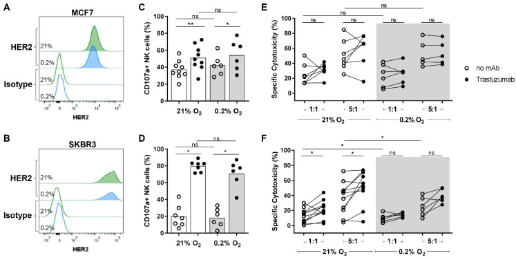
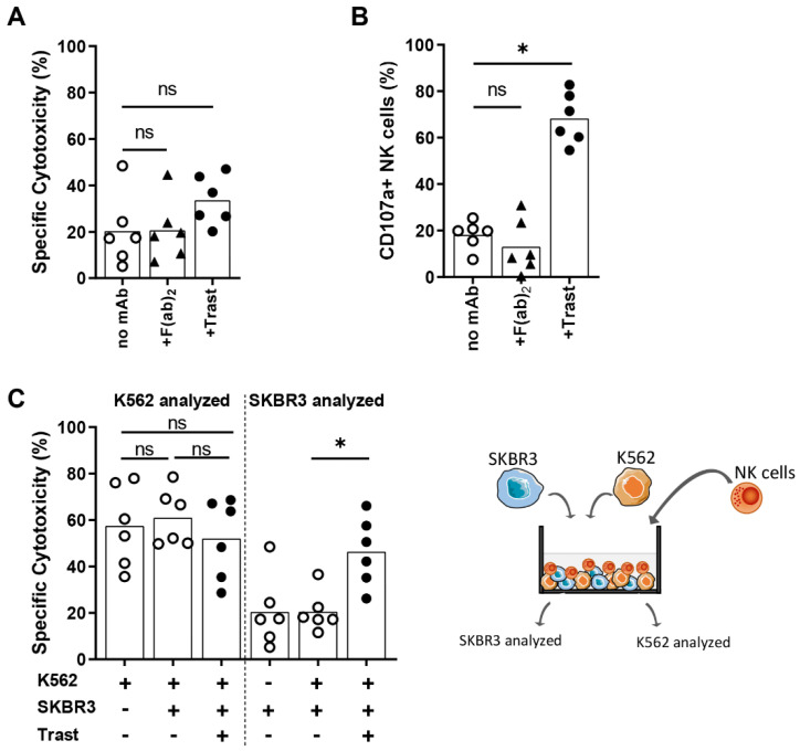
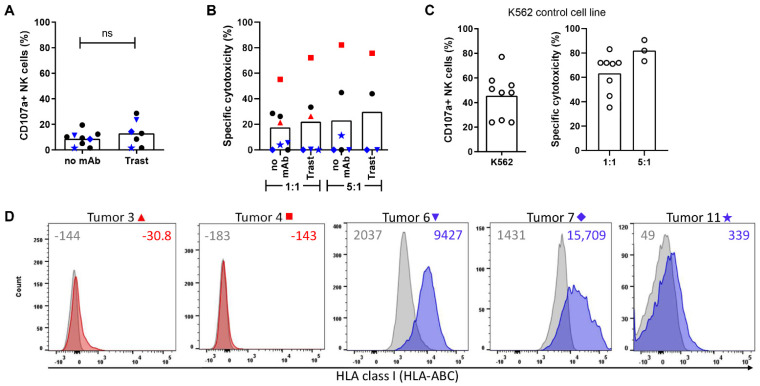
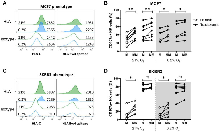
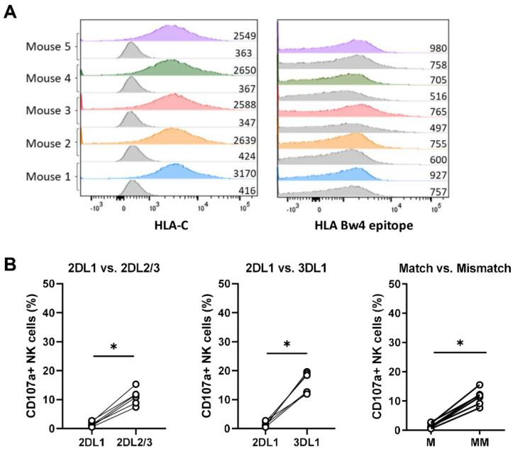
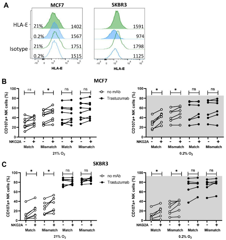
Natural killer (NK)-cell-based immunotherapies are an attractive treatment option for cancer. We previously showed that alloreactive mouse NK cells cured mice of 4T1 breast cancer. However, the tumor microenvironment can inhibit immune responses, and these suppressive factors must be overcome to unfold the NK cells' full anti-tumor potential. Here, we investigated the combination of antibody-dependent cellular cytotoxicity (ADDC) and the selection of KIR-HLA-ligand mismatched NK cells to enhance NK cell anti-breast cancer responses in clinically relevant settings. Donor-derived and IL-2-activated NK cells were co-cultured with patient-derived breast cancer cells or cell lines MCF7 or SKBR3 together with the anti-HER2 antibody trastuzumab. NK cells mediated anti-breast cancer cytotoxicity under normoxic and hypoxic conditions. Under both conditions, trastuzumab vigorously enhanced NK cell degranulation (CD107a) against HER2-overexpressing SKBR3 cells, but we observed a discrepancy between highly degranulating NK cells and a rather modest increase in cytotoxicity of SKBR3. Against patient-derived breast cancer cells, the anti-tumor efficacy was rather limited, and HLA class I expression seemed to contribute to inhibited NK cell functionality. KIR-ligand-mismatched NK cells degranulated stronger compared to the matched NK cells, further highlighting the role of HLA. In summary, trastuzumab and KIR-ligand-mismatched NK cells could be two strategies to potently enhance NK cell responses to breast cancer.

Keywords: HLA class I; alloreactive donor NK cells; antibody-dependent cellular cytotoxicity; breast cancer; tumor microenvironment.


Conflict of interest statement

G.M.J.B. is Chief Executive Officer/Chief Medical Officer/Cofounder of CiMaas, BV, Maastricht, The Netherlands. CiMaas is producing an ex vivo expanded NK cell product that will be used to treat myeloma patients.

Figures

Figure 1
Figure 1
NK cell efficacy against non-amplified and HER2 amplified cell lines with or without trastuzumab under normoxia (21% O2) and hypoxia (0.2% O2). (A,B) HER2 surface expression levels were determined by flow cytometry on non-amplified MCF7 cells (A) and HER2-amplified SKBR3 cells (B). (CF) For both degranulation (CD107a) and cytotoxicity assays, IL-2-activated NK cells were co-cultured with the target cells MCF7 or SKBR3 for 4 h either with 21% O2 or 0.2% O2 and analyzed by flow cytometry. CD107a assays were performed in 1:1 E:T ratios and % of CD107a+ NK cells are shown per donor against MCF7 (C) or SKBR3 (D) with the bar height indicating the mean. Cytotoxicity assays were done in 1:1 or 5:1 E:T ratios and dead target cells are shown as % of specific cytotoxicity for MCF7 (E) and SKBR3 (F). The donors from Figure 2 are included in the conditions with 21% O2 of (F). Each dot represents the average of duplicates from one NK cell donor. * p < 0.05, ** p < 0.01, ns = not significant.
Figure 2
Figure 2
NK cell efficacy against the target cells SKBR3 in the presence of an F(ab)2 fragment of trastuzumab. SKBR3 and K562 target cells were labeled with different dyes and co-cultured with IL-2-activated NK cells in a 1:1 E:T ratio with trastuzumab or with a F(ab)2 fragment of trastuzumab for 4 h with 21% O2. Flow cytometry was used to analyze specific cytotoxicity of tumor cells (A,C) and, in separate assays, degranulation of NK cells (B). (C) Target cells K562 and SKBR3 were combined in one well with or without trastuzumab and analyzed for specific cytotoxicity of K562 and SKBR3. The schematic setup is depicted on the right, and the full gating strategy is shown in Figure S3. Each dot represents one NK cell donor, and the average of duplicates is shown per donor with the bars indicating the mean. Data from three donors of (A) are also used in Figure 1F. * p < 0.05, ns = not significant.
Figure 3
Figure 3
NK cell degranulation and cytotoxic potential against HER2 non-amplified primary breast cancer. (A,B) Patient-derived breast cancer cells were dissociated to single cells and incubated with or without trastuzumab for 30 min and subsequently co-cultured with IL-2-activated NK cells in 1:1 or 5:1 E:T ratios for 16 h at 21% O2 and analyzed by flow cytometry. (A) Degranulating NK cells are shown as percentage CD107a+ NK cells, and each symbol (black and blue) represents one tumor sample; the blue symbols correspond to the colors of the histograms in (D). (B) Dead tumor cells are shown as specific cytotoxicity. The bar graphs indicate the mean, while each dot represents an individual breast cancer specimen. The red and blue symbols correspond to the histograms in (D). (C) K562 cells were included as a control in CD107a and cytotoxicity assays to show that NK cells were potent killers. (D) Histograms show HLA class I staining of breast cancer cells (red for samples with kill, blue for samples with low kill), and grey histograms show isotype controls. Tumors 3 and 4 (red symbols) were not available for CD107a assays in A. Median fluorescent intensity is depicted for each staining. ns = not significant.
Figure 4
Figure 4
NK cell degranulation of KIR-ligand-matched and KIR-ligand-mismatched NK cells in response to breast cancer cells under normoxia or hypoxia with or without trastuzumab. (A,B) MCF7 and SKBR3 cells were exposed to 21% O2 or 0.2% O2 for 16 h and stained for HLA surface expression by flow cytometry. Histograms from normoxic conditions are indicated in green, with hypoxic conditions in blue. (C,D) Degranulation assays were performed by co-culturing NK wells either with MCF7 or SKBR3 target cells with or without trastuzumab for 4 h at 21% O2 or 0.2% O2. Based on the KIR expression of NK cells and HLA expression of target cells, KIR-ligand-matched (M) and -mismatched (MM) NK cell subsets were analyzed and the percentage of degranulating NK cells is shown per subset as % CD107a+ NK cells. Each dot represents the average of duplicates from one NK cell donor. * p < 0.05, ** p < 0.01, ns = not significant.
Figure 5
Figure 5
NK cell degranulation of KIR-ligand-matched and -mismatched NK cells in response to MCF7 grown in vivo. MCF7 tumors that were grown in mice were harvested and dissociated into single-cell suspension before performing HLA staining with n = 5 tumors (A) or 4 h CD107a assays with n = 6 tumors (B). Within the CD107a assay, degranulation is shown for each receptor (left and middle graph) and also as KIR-ligand-matched (M, 2DL1) vs. -mismatched (MM, 2DL2/3 and 3DL1) NK cell subsets as % CD107a+ NK cells. Each dot represents a tumor isolated from one mouse and the average of duplicates is depicted. * p < 0.05.
Figure 6
Figure 6
NK cell degranulation of the NKG2A+ subsets in comparison to the NKG2A subsets is slightly enhanced. (A) HLA-E expression of MCF7 and SKBR3 was determined by flow cytometry. (B,C) For degranulation assays, NK cells were co-cultured either with MCF7 or SKBR3 targets cells with or without trastuzumab for 4 h at 21%O2 or 0.2% O2. By flow cytometry analysis, NK cells were grouped in NKG2A and NKG2A+ subsets (indicated by − and + below graphs) and further divided into KIR-ligand-matched and -mismatched subsets based on their KIR expression. For each subset, NK cell degranulation (CD107a in %) is depicted in response to MCF7 (B) or SKBR3 (C). Each dot represents one NK cell donor and the average of duplicates. * p < 0.05, ns = not significant.

References

    1. Torre L.A., Islami F., Siegel R.L., Ward E.M., Jemal A. Global cancer in women: Burden and trends. Cancer Epidemiol. Prev. Biomark. 2017;26:444–457. doi: 10.1158/1055-9965.EPI-16-0858. - DOI - PubMed
    1. Waks A.G., Winer E.P. Breast cancer treatment: A review. JAMA. 2019;321:288–300. doi: 10.1001/jama.2018.19323. - DOI - PubMed
    1. Wolff A.C., Hammond M.E.H., Allison K.H., Harvey B.E., Mangu P.B., Bartlett J.M., Bilous M., Ellis I.O., Fitzgibbons P., Hanna W. Human epidermal growth factor receptor 2 testing in breast cancer: American Society of Clinical Oncology/College of American Pathologists clinical practice guideline focused update. Arch. Pathol. Lab. Med. 2018;142:1364–1382. doi: 10.5858/arpa.2018-0902-SA. - DOI - PubMed
    1. Nahta R., Esteva F.J. Herceptin: Mechanisms of action and resistance. Cancer Lett. 2006;232:123–138. doi: 10.1016/j.canlet.2005.01.041. - DOI - PubMed
    1. Shimasaki N., Jain A., Campana D. NK cells for cancer immunotherapy. Nat. Rev. Drug Discov. 2020;19:200–218. doi: 10.1038/s41573-019-0052-1. - DOI - PubMed