. 2021 Jun 15;9(6):651.
doi: 10.3390/vaccines9060651.

Development of a Modular Vaccine Platform for Multimeric Antigen Display Using an Orthobunyavirus Model

Affiliations

Development of a Modular Vaccine Platform for Multimeric Antigen Display Using an Orthobunyavirus Model

Andrea Aebischer et al. Vaccines (Basel). .

Abstract

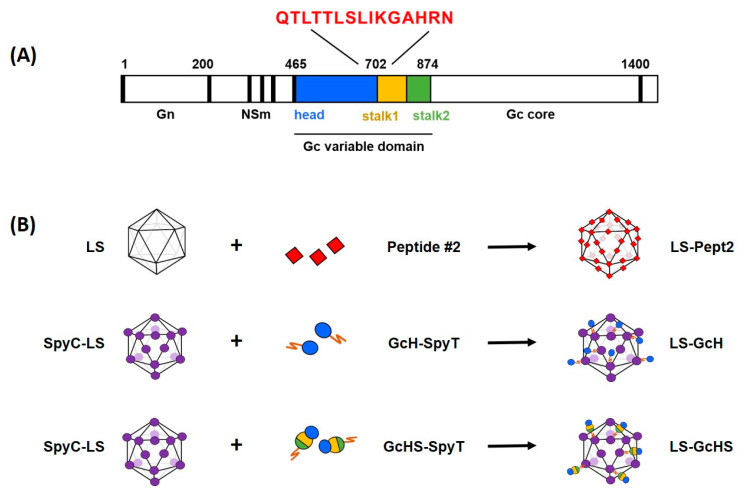
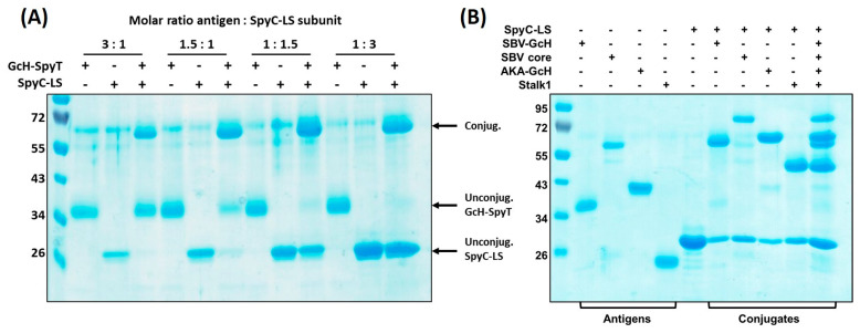
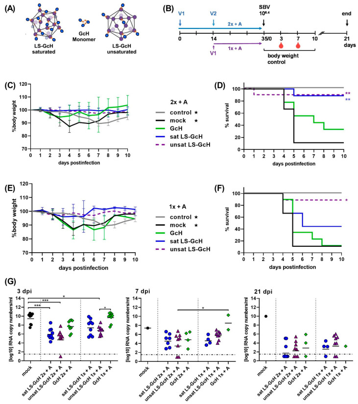
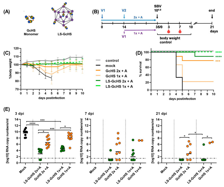
Emerging infectious diseases represent an increasing threat to human and animal health. Therefore, safe and effective vaccines that could be available within a short time frame after an outbreak are required for adequate prevention and control. Here, we developed a robust and versatile self-assembling multimeric protein scaffold particle (MPSP) vaccine platform using lumazine synthase (LS) from Aquifex aeolicus. This scaffold allowed the presentation of peptide epitopes by genetic fusion as well as the presentation of large antigens by bacterial superglue-based conjugation to the pre-assembled particle. Using the orthobunyavirus model Schmallenberg virus (SBV) we designed MPSPs presenting major immunogens of SBV and assessed their efficacy in a mouse model as well as in cattle, a target species of SBV. All prototype vaccines conferred protection from viral challenge infection and the multivalent presentation of the selected antigens on the MPSP markedly improved their immunogenicity compared to the monomeric subunits. Even a single shot vaccination protected about 80% of mice from an otherwise lethal dose of SBV. Most importantly, the MPSPs induced a virtually sterile immunity in cattle. Altogether, LS represents a promising platform for modular and rapid vaccine design.

Keywords: C1 production host; Schmallenberg virus; SpyCatcher/SpyTag; emerging infectious disease; epitope; lumazine synthase; modular vaccine; zoonosis.


Conflict of interest statement

Ronen Tchelet is an employee of Dyadic Netherlands BV. Jean-Christophe Audonnet is an employee of Boehringer Ingelheim Animal Health France. This does not alter the adherence to the Vaccines policies on sharing data.

Figures

Figure 1
Figure 1
Design and generation of vaccine candidates. (A) Schematic presentation of the SBV M-segment showing the selected model antigens within the variable N-terminal region of the Gc ectodomain: Gc head (GcH, aa 465–702), Gc head-stalk (GcHS, aa 465–874) and peptide epitope QTLTTLSLIKGAHRN (Pept2, aa 694–708); (B) Illustration of the applied vaccine design strategies and the resulting candidate vaccines. Peptide2 was genetically fused into the LS MPSP and the GcH or GcHS domain equipped with a SpyT were conjugated to the SpyC-LS MPSP by spontaneous isopeptide bonding.
Figure 2
Figure 2
Versatility of the Plug-and-display LS platform. (A) Conjugation efficiency is dependent on the molar ratio of antigen input vs. provided SpyC-LS subunits. Conjugation reactions were performed using molar ratios of 3:1; 1.5:1; 1:1.5 and 1:3 (antigen:SpyC-LS subunits). Reducing SDS-PAGE and Coomassie staining was performed after incubation for 48 h at RT. Input of SpyT-antigen and SpyC-LS are loaded next to the lanes showing the conjugation products; (B) Different antigens can be efficiently conjugated. Conjugation reactions were performed with SpyT-equipped SBV-GcH, SBV Gc core, SBV stalk1 and AKAV-GcH. All antigens were either conjugated separately with SpyC-LS or all together in one single reaction. Reducing SDS PAGE and Coomassie staining was performed after 48 h incubation at RT.
Figure 3
Figure 3
Final vaccine candidates. (A) Negatively stained TEM of the SpyC-LS MPSP. (B) Reducing SDS-PAGE and Coomassie staining showing input partners and the respective conjugation products of the saturated (antigen:LS subunit = 3:1) and unsaturated (1:1.5) LS-GcH MPSP preparations used for immunizations. Saturated LS-GcH particles were purified by dialysis in order to remove unconjugated GcH monomers as indicated by arrows. (C) Reducing SDS-PAGE and Coomassie staining showing input partners next to the conjugation product of the final LS-GcHS (antigen:LS subunits = 1:3) MPSP vaccine. (D) Reducing SDS-PAGE and Coomassie staining of the LS-MPSP after N-terminal genetic fusion of the peptide epitope #2 (LS-Pept2) in comparison to the SpyC-LS backbone. (E) TEM images of the finally selected vaccine candidates for evaluation in the target species. From left to right: LS-Pept2, LS-GcH saturated, LS-GcH unsaturated, LS-GcHS. Scale bars 200 nm.
Figure 4
Figure 4
IFNAR-/- mouse trial #1. Efficacy of the LS-conjugated GcH domain compared to the monomeric GcH and the LS-presented peptide epitope QTLTTLSLIKGAHRN. (A) Schematic of vaccines used for the immunizations in this trial; (B) Experimental scheduling. V1 and V2 indicate time-points of the 1st and 2nd vaccination; (C) Body weight development after challenge infection in groups vaccinated twice with adjuvanted vaccines (2x + A). Each line represents the mean value of the respective group with standard deviation (SD); (D) Survival curves in groups vaccinated 2x + A; (E) Body weight development after challenge infection in groups vaccinated once with adjuvanted vaccines (1x + A) or twice in the absence of adjuvant (2x w/o A); (F) Survival curves in groups vaccinated 1x A or 2x w/o A; For (C,E) as well as for (D,F) data of the respective control and mock groups (marked with stars) were inserted in both graphs; (G) SBV RNA detected by RT-qPCR in EDTA blood samples of surviving animals in each group at 3, 7 or 21 dpi, respectively. Dashed lines indicate the detection limit of the RT-qPCR assay. Samples of animals that succumbed to infection or had to be euthanized prior to or on the respective sampling day were not included. In (G) statistical analysis was performed using the Kruskal–Wallis test followed by Dunn´s test for comparisons between individual groups. p values < 0.05 were considered significant. (* p < 0.05; ** p < 0.01; *** p < 0.001). Only significant differences between groups are labeled. Differences that are not significant (p > 0.05) are not separately indicated. In (D,F) significant differences compared to the mock control were calculated using the Mantel–Cox test (* 0.0332; ** 0.0021; *** 0.0002; **** <0.0001). Comparisons between all groups against each other are not indicated but are shown in Table S2.
Figure 5
Figure 5
IFNAR-/- mouse trial #2. Efficacy of saturated and unsaturated LS-GcH MPSPs in comparison to monomeric GcH. (A) Schematic of vaccines used for the immunizations in this trial; (B) Experimental scheduling. V1 and V2 indicate the time-points of the 1st and 2nd vaccination; (C) Body weight development after challenge infection in groups vaccinated twice with adjuvanted vaccines (2x + A). Each line represents he mean value of the respective group with SD; (D) Survival curves in groups vaccinated 2x + A; (E) Body weight development after challenge infection in groups vaccinated once with adjuvanted vaccines (1x + A); (F) Survival curves in groups vaccinated 1x + A; For (C,E) as well as for (D,F) data of the respective control and mock groups (marked with stars) were inserted in both graphs; (G) SBV RNA detected by RT-qPCR in EDTA blood samples of surviving animals in each group at 3, 7 or 21 dpi, respectively. Dashed lines indicate the detection limit of the RT-qPCR assay. Samples of animals that succumbed to infection or had to be euthanized prior to or on the respective sampling day were not included. In (G) Statistical analysis was performed using the Kruskal–Wallis test followed by Dunn´s test for comparisons between individual groups. p values < 0.05 were considered significant. (* p < 0.05; ** p < 0.01; *** p < 0.001). Only significant differences between groups are labeled. Differences that are not significant (p > 0.05) are not separately indicated. In (D,F) significant differences compared to the mock control were calculated using the Mantel–Cox test (* 0.0332; ** 0.0021; *** 0.0002; **** <0.0001). Comparisons between all groups against each other are not indicated but are shown in Table S2.
Figure 6
Figure 6
IFNAR-/- mouse trial #3. Evaluation of the C1-produced GcHS domain and the protective efficacy of the monomeric and LS-conjugated antigen. (A) Schematic of vaccines used for the immunizations in this trial; (B) Experimental scheduling; (C) Body weight development after challenge infection, each line represents the mean value of the respective group with SD; (D) Survival curves after challenge infection. One animal in group LS-GcHS vaccinated twice (2x + A) accidentally died during blood sampling in a manner unrelated to SBV infection; (E) SBV RNA detected by RT-qPCR in EDTA blood samples of surviving animals in each group at 3, 7 or 21 dpi, respectively. Dashed lines indicate the detection limit of the RT-qPCR assay. Samples of animals that succumbed to infection or had to be euthanized prior to or on the respective sampling day were not included. In (E) statistical analysis was performed using the Kruskal–Wallis test followed by Dunn´s test for comparisons between individual groups. p values < 0.05 were considered significant (* p < 0.05; ** p < 0.01; *** p < 0.001; **** p < 0.0001). Only significant differences between groups are labeled. Differences that are not significant (p > 0.05) are not separately indicated. In (D) significant differences compared to the mock control were calculated using the Mantel–Cox test (* 0.0332; ** 0.0021; *** 0.0002; **** <0.0001). Comparisons between all groups against each other are not indicated but are shown in Table S2.
Figure 7
Figure 7
Evaluation of finally selected vaccine candidates in cattle. (A) Schematic illustration of the final candidate vaccines used in the trial; (B) Vaccination and sampling schedule, V1 and V2 indicate time-points of the 1st and 2nd immunization; (C) Mean body temperatures after vaccination and challenge infection. Each line represents the mean value of the respective groups with SD; (D) Development of neutralizing antibodies after immunizations and challenge infection, determined by VNT. Mean values and SD of ND50 titers are indicated for each group; (E) Detection of SBV-Nucleoprotein-specific antibodies by ELISA in serum samples collected on 21 dpi; (F) SBV genome equivalents in serum samples that were collected daily for 10 days after challenge infection. Geometric means are indicated by connected solid lines; (G) SBV RNA detected in tissue samples of each animal collected at necropsy 28 dpi. In (F,G) dashed lines indicate the detection limit of the RT-qPCR assay. In (DG) one animal vaccinated with LS-Pept2 was excluded from data analysis, since it showed no reaction to either immunization or challenge infection. No SBV RNA was found in any sample tested by RT-qPCR and the animal did not develop antibodies towards the LS-Pept2 antigen and neither neutralizing nor N-specific antibodies after challenge infection.

References

    1. Hassell J.M., Begon M., Ward M.J., Fèvre E.M. Urbanization and Disease Emergence: Dynamics at the Wildlife-Livestock-Human Interface. Trends Ecol. Evol. 2017;32:55–67. doi: 10.1016/j.tree.2016.09.012. - DOI - PMC - PubMed
    1. Jones K.E., Patel N.G., Levy M.A., Storeygard A., Balk D., Gittleman J.L., Daszak P. Global trends in emerging infectious diseases. Nature. 2008;451:990. doi: 10.1038/nature06536. - DOI - PMC - PubMed
    1. Hemida M.G., Chu D.K., Poon L.L., Perera R.A., Alhammadi M.A., Ng H.Y., Siu L.Y., Guan Y., Alnaeem A., Peiris M. MERS coronavirus in dromedary camel herd, Saudi Arabia. Emerg. Infect. Dis. 2014;20:1231–1234. doi: 10.3201/eid2007.140571. - DOI - PMC - PubMed
    1. Zanluca C., dos Santos C.N.D. Zika virus—An overview. Microbes Infect. 2016;18:295–301. doi: 10.1016/j.micinf.2016.03.003. - DOI - PubMed
    1. Wang C., Horby P.W., Hayden F.G., Gao G.F. A novel coronavirus outbreak of global health concern. Lancet. 2020;395:470–473. doi: 10.1016/S0140-6736(20)30185-9. - DOI - PMC - PubMed

LinkOut - more resources