Review
. 2021 Jun 15;10(12):2639.
doi: 10.3390/jcm10122639.

Applications and Limitations of Neuro-Monitoring in Paediatric Anaesthesia and Intravenous Anaesthesia: A Narrative Review

Affiliations
Review

Applications and Limitations of Neuro-Monitoring in Paediatric Anaesthesia and Intravenous Anaesthesia: A Narrative Review

Chiara Grasso et al. J Clin Med. .

Abstract

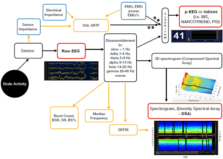
Safe management of anaesthesia in children has been one of the top areas of research over the last decade. After the large volume of articles which focused on the putative neurotoxic effect of anaesthetic agents on the developing brain, the attention and research efforts shifted toward prevention and treatment of critical events and the importance of peri-anaesthetic haemodynamic stability to prevent negative neurological outcomes. Safetots.org is an international initiative aiming at raising the attention on the relevance of a high-quality anaesthesia in children undergoing surgical and non-surgical procedures to guarantee a favourable outcome. Children might experience hemodynamic instability for many reasons, and how the range of normality within brain autoregulation is maintained is still unknown. Neuro-monitoring can guide anaesthesia providers in delivering optimal anaesthetic drugs dosages and also correcting underling conditions that can negatively affect the neurological outcome. In particular, it is referred to EEG-based monitoring and monitoring for brain oxygenation.

Keywords: EEG-derived monitor; anaesthesia; brain oxygenation; depth of anaesthesia; near-infrared-spectroscopy; neuromonitoring; paediatric.


Conflict of interest statement

The authors declare no conflict of interest.

Figures

Figure 1
Figure 1
Mechanism of action of the currently available EEG derived anaesthesia monitoring (in red). The sensor is placed on the forehead and records the electrical potentials produced in the cortex resulting in the raw EEG. The majority of the EEG-derived monitors work by disassembling a complex EEG waveform (the raw EEG), into many series of waves of different frequencies (Hertz—Hz). These, together with several of the EEG variables, are converted into a single index through mathematical algorithms by the processed EEG (p-EEG) devices. The obtained index represents the level of hypnosis. However, the index can also be affected by the Electromyography (EMG) and the Signal Quality Index (SQI or ARTF). The spectrogram, is a real time monitoring which portrays all the EEG frequencies and their power over the time in a three-dimensional method (3D spectrogram or Compressed Spectral Array). The latter is then integrated in a two-dimensional plot using colours to represent different powers in the Density Spectral Array (DSA). Derived additional parameters are the Spectral Edge Frequency (SEF95), Median Frequency and the Burst Count (burst/minute) Suppression Rate (SR), Burst Suppression Ratio (BSR), Suppression Ratio (SR) or BS% Indicator (Burst suppression percentage). These can be either represented on the display by a number or visualized on the Spectrogram.
Figure 2
Figure 2
Cerebral oxygen delivery and consumption balance and contributing factors to cerebral oxygen saturation during anaesthesia. HR, heart rate; BP, blood pressure; SatO2, oxygen saturation; CO2, carbon dioxide; T°, temperature; ScO2 cerebral oxygen saturation.

References

    1. Cornelissen L., Kim S.E., Lee J.M., Brown E.N., Purdon P.L., Berde C.B. Electroencephalographic markers of brain development during sevoflurane anaesthesia in children up to 3 years old. Br. J. Anaesth. 2018;120:1274–1286. doi: 10.1016/j.bja.2018.01.037. - DOI - PMC - PubMed
    1. Valkenburg A.J., De Leeuw T.G., Tibboel D., Weber F. Lower bispectral index values in children who are intellectually disabled. Anaest. Analg. 2009;109:1428–1433. doi: 10.1213/01ANE.0b013e3181ba43b2. - DOI - PubMed
    1. Brandt S.P., Walsh E.C., Cornelissen L., Lee J.M., Berde C., Shank E.S., Purdon P.L. Case Studies Using the Electroencephalogram to Monitor Anesthesia-Induced Brain States in Children. Anesth. Analg. 2020;131:1043–1056. doi: 10.1213/ANE.0000000000004817. - DOI - PMC - PubMed
    1. Walsh E.C., Lee J.M., Terzakis K., Zhou D.W., Burns S., Buie T.M., Firth P.G., Shank E.S., Houle T.T., Brown E.N. Age-dependent changes in the propofol-induced electroencephalogram in children with autism spectrum disorder. Front. Syst. Neurosci. 2018;12:23. doi: 10.3389/fnsys.2018.00023. - DOI - PMC - PubMed
    1. Rigouzzo A., Khoy-Ear L., Laude D., Louvet N., Moutard M.-L., Sabourdin N., Constant I. EEG profiles during general anesthesia in children: A comparative study between sevoflurane and propofol. Paediatr. Anaesth. 2019;29:250–257. doi: 10.1111/pan.13579. - DOI - PubMed

LinkOut - more resources