Review
. 2021 Jun 6;10(6):1413.
doi: 10.3390/cells10061413.

Urine-Derived Epithelial Cells as Models for Genetic Kidney Diseases

Affiliations
Review

Urine-Derived Epithelial Cells as Models for Genetic Kidney Diseases

Tjessa Bondue et al. Cells. .

Abstract

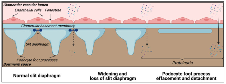
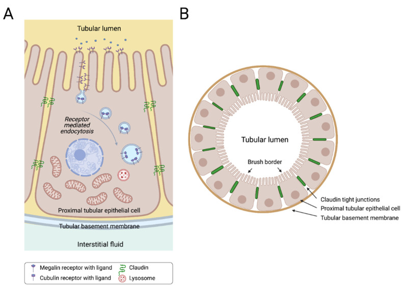
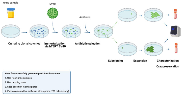
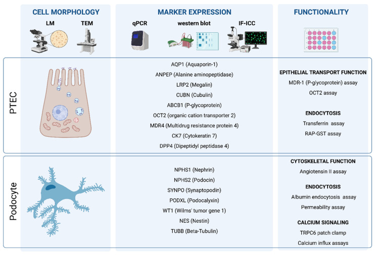
Epithelial cells exfoliated in human urine can include cells anywhere from the urinary tract and kidneys; however, podocytes and proximal tubular epithelial cells (PTECs) are by far the most relevant cell types for the study of genetic kidney diseases. When maintained in vitro, they have been proven extremely valuable for discovering disease mechanisms and for the development of new therapies. Furthermore, cultured patient cells can individually represent their human sources and their specific variants for personalized medicine studies, which are recently gaining much interest. In this review, we summarize the methodology for establishing human podocyte and PTEC cell lines from urine and highlight their importance as kidney disease cell models. We explore the well-established and recent techniques of cell isolation, quantification, immortalization and characterization, and we describe their current and future applications.

Keywords: Fanconi syndrome; PTECs; glomerular diseases; inherited renal disorders; kidney disease cellular models; podocytes; proximal tubular epithelial cells; renal tubular acidosis; urine-derived cells.


Conflict of interest statement

The authors declare no conflict of interest.

Figures

Figure 1
Figure 1
Podocytes: key element of the glomerular filtration barrier. Podocytes are highly differentiated cells with a specific cellular architecture, comprising primary and secondary foot processes that wrap around the glomerular capillaries, and express essential proteins that define the slit diaphragm and permselectivity of the glomerular filtration barrier. Kidney diseases affecting the podocytes (podocytopathies) can be characterized by the loss of the slit diaphragm, podocyte foot process effacement and detachment, which clinically manifests as glomerular proteinuria.
Figure 2
Figure 2
Sagittal (A) and cross (B) sections of a human proximal tubule. Proximal tubular epithelial cells (PTECs) are indispensable cells for the nephron tubular system. PTECs are highly metabolically active cells that are responsible for reabsorbing many of the filtered solutes in the proximal part of the nephron. These cells are typically characterized by a brush border at the apical side, and high numbers of mitochondria at the basal side of the cell. Megalin and cubulin are characteristic multi-ligand receptors for PTECS, located at the brush border, responsible for receptor-mediated endocytosis for various ligands.
Figure 3
Figure 3
Procedure of isolation, immortalization and subcloning of urine-derived kidney epithelial cells. Fresh urine is cultured in a specific medium that selects for specific types of kidney epithelial cells at 37 °C. Immortalization is performed by use of SV40 and the hTERT gene, upon which antibiotic selection is used to isolate the successfully immortalized cells. Clonal colonies are selected, in order to isolate and expand specific cell clones that will be characterized by biomarker expression and stored for cryopreservation.
Figure 4
Figure 4
Technology toolbox for characterization of urine-derived kidney epithelial cells. Urine-derived kidney epithelial cells are characterized based on their cellular morphology, the expression of cell-type specific markers, and their functionality via cell-specific assays. To this end, microscopy, including light microscopy (LM) and transmission electron microscopy (TEM), qPCR and Western Blot plus immunocytochemistry are useful technologies.

References

    1. Sutherland G.R., Bain A.D. Culture of cells from the urine of newborn children. Nature. 1972;239:231. doi: 10.1038/239231a0. - DOI - PubMed
    1. Linder D. Culture of cells from the urine and bladder washings of adults. Somatic Cell Genet. 1976;2:281–283. doi: 10.1007/BF01538966. - DOI - PubMed
    1. Felix J.S., Sun T.T., Littlefield J.W. Human epithelial cells cultured from urine: Growth properties and keratin staining. Vitr. J. Tissue Cult. Assoc. 1980;16:866–874. doi: 10.1007/BF02619424. - DOI - PubMed
    1. Elliott A.Y., Bronson D.L., Stein N., Fraley E.E. In Vitro Cultivation of Epithelial Cells Derived from Tumors of the Human Urinary Tract. Cancer Res. 1976;36:365–369. - PubMed
    1. Elliott A.Y., Bronson D.L., Cervenka J., Stein N., Fraley E.E. Properties of Cell Lines Established from Transitional Cell Cancers of the Human Urinary Tract. Cancer Res. 1977;37:1279–1289. - PubMed

MeSH terms